Open Access, Peer-reviewed
eISSN 2093-9752
Open Access, Peer-reviewed
eISSN 2093-9752
Young-Tae Lim
Jun Sung Park
Jae Woo Lee
Moon-Seok Kwon
http://dx.doi.org/10.5103/KJSB.2017.27.2.125 Epub 2017 July 15
Abstract
Objective: The aim of this study was to investigate the effect of targeted knee flexion angle on biomechanical factors of knee joint between upward and downward phases during the forward lunge.
Method: Eight elderly subjects (age: 22.23±1.51 years, weight: 69±6.63 kg, height: 174.88±6.85 cm) participated in this study. All reflective marker data and ground reaction force during a forward lunge were collected. The knee joint movement and reaction force and joint moment at maximum knee flexion angle were compared by repeated measures one-way analysis of variance (ANOVA) (p<.05). The peak knee joint reaction force and joint moment between upward and downward phases were compared by repeated measures two-way ANOVA (p<.05).
Results: The anterior and vertical knee joint movements, reaction force, and extensor moment of 80° targeted knee flexion condition at maximum knee flexion angle was greater than both 90° and 100° conditions (p<.05). The 80° knee flexed angle condition had greater peak joint reaction force and extensor moment compared with both 90° and 100° conditions between upward and downward phases during the forward lunge.
Conclusion: As the targeted knee joint flexion angle increases, knee joint movement and kinetic variables become greater during the forward lunge exercise.
Keywords
Forward lunge Targeted knee flexion angle Joint reaction force Joint moment
Forward lunge is one of the motions often performed for daily physical activities, such as climbing stairs and hiking, as well as during various sports activities and physical muscle training (Hofmann, Holyoak, & Juris, 2017; Kong, 2014; Kritz, Cronin, & Hume, 2009). A forward lunge refers to a motion wherein one foot is moved in front of the other in the starting position, and then the upper torso is moved downward from the point of the foot, contacting the ground surface to the targeted knee being flexed, after which the upper torso is moved upward to return to the starting position (Escamilla et al., 2010; Hofmann et al., 2017; Kritz et al., 2009). The upward-downward movement of the upper torso during a forward lunge affects the continuous eccentric flexion and concentric extension performed by the musculoskeletal system of the legs. In addition, positional change by the center of gravity or increased external weight load has been reported to increase the contractile force in the lower extremity muscles that support weight and core muscles used to maintain physical balance (Hofmann et al., 2017; Park, Lee, & Choi, 2010). Because of these reasons, forward lunge has positive aspects for increasing muscle strength and endurance in lower extremity and core muscles and for rehabilitation training after knee surgery (Escamilla et al., 2010; Hall et al., 2015). However, because forward lunge also generates a significant amount of weight loading together with knee flexion in the front leg, many previous studies have been conducted on injury factors in the knee during the forward lunge (Escamilla et al., 2008; Escamilla et al., 2010; Farrokhi et al., 2008).
Escamilla et al. (2010) categorized forward lunge as a lunge with a stride where the lunge motion is performed after taking a step forward, and the lunge without a stride where both feet are positioned stably in a lunge position, and the actual lunge is performed as an up-down motion. The lunge with a stride has been reported to require high skill level for achieving the necessary lower extremity muscle strength and physical stability. The forward lunge with a stride is form of a typical closed kinetic chain exercise (CKCE) for training lower extremity muscles, and because it involves moving one foot forward to contact the ground surface, while transferring the weight to that front leg (Heijne et al., 2004; Park et al., 2010), the anterior-posterior (AP) and lateral motions in the knee joint in the front leg become restricted during the downward phase (Kritz et al., 2009). Particularly, increase in the amount of load exerted on the knee joint and combined contraction of the quadriceps, biceps femoris, and hip abductor and adductor muscles at the point of maximum knee flexion have an impact on the kinetic factors of the knee joint (Ekstrom, Donatelli, & Carp, 2007; Henriksen, Alkjaer, Simonsen, & Bliddal, 2009; Hofmann et al., 2017).
Stride length and trunk position factors have been reported to affect peak reaction force and moment generated in the knee of the front leg during forward lunge (Escamilla et al., 2008; Escamilla et al., 2010; Farrokhi et al., 2008). Previous studies on knee joint loading during body movement have reported that excessive knee extension and abduction and tibial external rotation during weight-bearing motion may increase the risk of anterior cruciate ligament (ACL) injury (Boden, Dean, Feagin, & Garrett, 2000; Hewett, Myer, & Ford, 2006; Kim & Youm, 2013). In addition, forward lunge, a form of CKCE, is recognized as having the potential for causing knee injury due to factors associated with excessive abduction and external rotation moments for maintaining lateral balance and knee reaction force that undergoes changes from knee flexion (Escamilla et al., 2010; Kong, 2014; Kritz et al., 2009; Park et al., 2010). Approximately 70% of ACL injuries that occur in the knee during sports activities are reported as non-contact injuries that occur during deceleration (Fleming et al., 2001; Hewett et al., 2005; Hewett et al., 2006; Jin, 2013; Kwon, 2012).
The knee reaction force and moment that are generated when per- forming knee flexion together with weight-bearing exercise have been reported as factors that can cause ACL injury (Fleming et al., 2001; Markolf et al., 1995). Increased knee flexion angle during a forward lunge causes forward movement of upper body segment and trunk segment that have the highest weight ratio to generate a large reaction force on the ground surface contacted by the front foot (Escamilla et al., 2010; Henriksen et al., 2009). Moreover, AP and lateral positional changes in the knee based on the knee flexion angle in the front leg increase the contractile force of the muscles involved in the rotational motion of the knee, which have been reported to have an impact on increased tibial anterior reaction force and risk of ACL injury (Arms et al., 1984; Escamilla et al., 2010; Li et al., 2004). Because of these reasons, it is believed that knee flexion angle in the downward phase should be considered to prevent injuries to the knee joints during training with forward lunge motion for muscle strengthening and rehabilitation training.
Accordingly, the objective of the present study was to analyze the effects of changes in knee flexion angle of the front leg during a forward lunge on the kinetic factors. For this objective, the flexion angle in the targeted knee was set to 80°, 90°, and 100°, and the range of motion, reaction force and moment in the targeted knee at various points of knee flexion angle were analyzed, and the peak reaction force and moment in the knee generated in upward and downward phases were also analyzed.
1. Participants
The participants in the present study consisted of eight male undergraduate physical education majors (age: 22.23±1.51 years, weight: 69±6.63 kg, and height: 174.88±6.85 cm) who had at least one year of weight training; used the right leg as the dominant leg; and did not have any lower extremity joint injuries in the past one year. All candidates were provided the full explanation on the objective and procedures of the study, and those who consented to participate in the study were included.
2. Data collection
Height and weight were measured in all participants. Moreover, an anthropometer (SM-324) was used to measure body segment length and circumference that were required for applying the scaling method for body segment parameters (Zatsiorsky, Seluyanov, & Chugunova, 1990). The participants stood with their feet at shoulder width apart in their respective ready position marked horizontally on the floor, after which, they performed a forward lunge motion of moving the right foot forward. After the marking the position of the front foot at 90° knee flexion angle lunge on the floor, the participants were instructed to perform 80° and 100° lunges with the same stride length. To prevent injuries, the participants were provided adequate time for stretching. To reduce errors in knee flexion angle measurements under the three different conditions, a goniometer (HI-SXJDC01, Jakemy, Hong Kong) was used to measure the knee flexion angle at each condition before actually performing the forward lunge motions. For this, knee flexion angle was defined as the internal angle formed by the straight line from the lateral epicondyle to the greater trochanter and the straight line from the lateral epicondyle to the lateral malleolus, as measured by the goniometer. To familiarize the participants with the three targeted knee flexion angles, they were allowed to practice the forward lunge motions for 20 min. Image analysis was used to select motions that were performed within 5° of the three different targeted knee flexion angle conditions (Table 1). All participants were instructed to hold a 5-kg dumbbell in each hand to apply a 10-kg weight. Moreover, the participants were also instructed to fully extend the elbows during the forward lunge motion to minimize rotation of the shoulder joints, and the motions were performed while maintaining the upper body upright and facing forward to control any movement of the upper torso.
|
Targeted knee flexion angle Mean (SD) |
||
|
80° |
90° |
100° |
|
78.719 (4.24) |
89.313 (2.45) |
101.422 (3.36) |
After the practice period, all participants wore just black shorts made of spandex material for the experiment, and 35 15-mm reflective markers suitable for a plug-in-gait model were attached on the participants for 3D motion analysis during the forward lunge (Table 2). Before the lunge experiment, 3D coordinate values of the camera were generated from images acquired while waving a T-shaped wand with four reflective markers in space, after which, nonlinear transformation (NLT) type of calibration was performed. Subsequently, a global coordinate system was established with the Y-axis defined as the AP direction, which was the direction where the motion was performed; X-axis defined as the lateral direction; and Z-axis defined as the vertical direction. Using the reflective markers attached to the body, a local coordinate system for the head, torso, pelvis, thigh, lower leg, foot, upper arm, forearm, and hand segments was defined for calculation of segmental movements during the forward lunge (Figure 1).
|
1. Right, left toe on the shoes
2. Right,
left foot upper posterior calcaneus 3. Right, left lateral malleolus 4. Right, left medial
malleolus 5. Right, left lateral
tibia 6. Right, left lateral
epicondyle 7. Right, left medial
epicondyle 8. Right,
left lateral thigh (half way between ankle & knee) 9. Right, left greater
trochanter 10. Right and left
anterior superior iliac spine |
11. Middle of posterior superior iliac spine 12. Right, left 3rd metacarpal head 13. Right, left lateral aspect of head of ulnar 14. Right, left lateral aspect of head of radius 15. Right, left humeral lateral epicondyle 16. Right, left humeral medial epicondyle 17. Right,
left lateral acromion 18. Upper ridge of ear
19. Middle of the forehead 20. 7th cervical
vertebral, 12th thoracic vertebrae |
All participants performed three repetitions of forward lunge under each of the three knee flexion angle conditions of 80°, 90°, and 100°. Moreover, to minimize the phenomenon of musculoskeletal manipulation from cognition due to repeating the same motion under the same knee flexion angle condition, the angle conditions were selected randomly and relayed to the participants. The 3D motion data of the participants during forward lunge were collected using eight infrared motion-capture cameras (T10S, Vicon, UK) at 200 Hz, whereas two force platforms (OR6-7-1000, AMTI, Inc, Watertown, MA, UK, sampling rate: 2,000 Hz) were used to collect the ground reaction force (GRF) data (Table 3).
|
Equipment |
Model |
Country |
|
Motion capture |
MX-T10S |
Vicon (UK) |
|
Data acquisition |
Giganet |
Vicon (UK) |
|
Analysis software |
NEXUS 1.8.3 |
Vicon (UK) |
|
Kwon3D XP |
Visol (Korea) |
|
|
Goniometer |
HI-SXJDC01 |
Jakemy (Hong Kong) |
|
Force platform |
OR6-7-1000 |
AMTI (USA) |
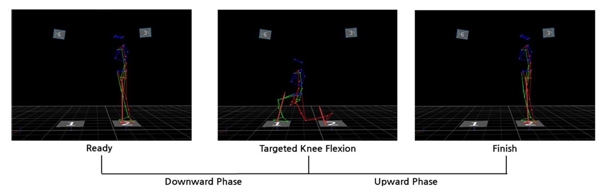
Figure 1 shows the forward lunge motion defined into three events for data analysis, which consisted of ready event, when the right heel was lifted off the ground; targeted knee flexion angle event, when the knee joint in the front leg was fully flexed; and finish event, when the front leg returned to its original position. Moreover, the motion was also defined into two phases: downward phase from the ready event to the targeted knee flexion angle event when the upper torso moved towards the ground and upward phase from the targeted knee flexion angle event to the finish event, when the upper torso moved away from the ground.
The GRF data and 3D data of biomarkers collected during the for- ward lunge were synchronized via Giganet (Vicon, UK) and saved to a computer, after which, Vicon Nexus Version 1.8 (Vicon, UK) was used for signal processing. Here, the data from the reflective markers attached on the body were filtered via fourth-order Butterworth filter (8 Hz), and the GRF data were filtered via fourth-order low-pass Butterworth filter (50 Hz), and then, saved as C3D files. The forward lunge C3D data saved in Vicon Nexus program was imported into Kwon3d XP software (Visol Inc, Korea), and the data were converted. After establishing a local coordinate system (X-axis: lateral direction; Y-axis: AP direction, and Z-axis: long axis of the segments) for the body segments with Kwon3d body modeling, the joint angle was calculated using the Cardan orientation method that calculates the relative orientation angles of lower leg segment coordinates to the thigh segment coordinates. Moreover, knee joint movement was measured by calculating the lateral, AP, and vertical range of motion from the point the front foot touched the ground to the point of reaching the maximum knee flexion (Table 4). The inertia values of body segments during the forward lunge by the participants, as well as kinematic and GRF data obtained through the experiment, were substituted into an inverse dynamics method to calculate the reaction force and moment factors generated in the knee joint (Kong, 2014). Moreover, reaction forces generated in three axes of the knee were defined as right (+)/left (-), anterior (+)/posterior (-), and up (+)/down (-). The knee joint moment was defined as the following: positive (+) and negative (-) values in the lateral axis as exten- sion and flexion, respectively; in the AP axis as adduction and abduction, respectively; and in the vertical axis as internal and external rotation, respectively. The values for knee joint reaction force and moments generated at the point of targeted knee flexion angle during forward lunge were calculated based on three different knee flexion angle conditions. Moreover, the peak knee reaction force and moment at downward and upward phases, which separated the points before and after the targeted knee flexion angle, were also calculated. All kinetic data were standardized by the weight of each participant.
|
Knee joint variables |
Targeted knee flexion mean (SD) |
F-value |
p-value |
|||
|
80° |
90° |
100° |
||||
|
Knee joint displacement (cm) |
Medial-lateral |
3.151 (2.84) |
2.955 (2.28) |
2.451 (2.01) |
.753 |
.489 |
|
Anterior-posterior |
40.984 (4.58)a,c |
30.057 (5.47)a,b |
24.511 (7.63)b,c |
31.341 |
.000 |
|
|
Vertical |
-6.641 (0.79)a,c |
-2.910 (2.28)a,b |
-0.244 (1.24)b,c |
60.335 |
.000 |
|
|
Knee joint reaction force (N/BW) |
Medial-lateral |
0.108 (0.16)c |
0.265 (0.33)b |
0.404 (0.30)b,c |
11.388 |
.001 |
|
Anterior–posterior |
0.693 (0.48)a,c |
1.397 (0.78)a,b |
1.940 (0.77)b,c |
21.800 |
.000 |
|
|
Vertical |
-8.956 (0.79)a,c |
-7.725 (0.84)a |
-5.640 (3.08)c |
5.810 |
.015 |
|
|
Knee joint moment (Nm/BW) |
Extensor |
0.980 (0.14)a,c |
0.717 (0.22)a,b |
0.362 (0.27)b,c |
26.584 |
.000 |
|
Abductor |
-0.223 (0.15) |
-0.241 (0.15) |
-0.182 (0.11) |
1.854 |
.193 |
|
|
External rotator |
-0.002 (0.01) |
-0.019 (0.03) |
-0.032 (0.05) |
2.719 |
.101 |
|
3. Statistical analysis
The mean value from three repetitions performed by each participant was used to test the differences in knee reaction force and moment at the point of targeted knee flexion angle based on the knee flexion angle during the forward lunge, and the differences in peak knee reaction force and moment generated downward and upward phases. The mean values of all variables were inputted into a statistics program (SPSS 18.0 SPSS Inc., Chicago, IL 60606, USA) based on knee flexion angles and phases. A repeated measures one-way analysis of variance (ANOVA) was used to analyze the differences between knee reaction force and moment factors at the point of targeted knee flexion angle during the forward lunge, whereas repeated measures two-way ANOVA was used to analyze the differences between downward and upward phases based on targeted knee flexion angles. A least significant difference (LSD) analysis was used for post-hoc test, with the significance level set to α=.05.
The results of the analysis on the kinetic factors during the forward lunge performed by the participants based on changes in the knee joint angles are shown in Table 4. As shown, knee joint measurement at the point of targeted knee flexion angle during the forward lunge showed statistically significant differences in the anterior (F=31.341, p<.05) and downward (F=60.335, p<.05) movements. The targeted knee flexion angle under the 80° condition showed significantly larger anterior and downward movements than under 90° and 100° conditions (p<.05).
Moreover, significant differences in the lateral (F=11.388, p<.05), anterior (F=21.800, p<.05), and vertical (F=5.810, p<.05) joint reaction forces of the knee based on targeted knee flexion angle conditions. Under the 100° flexion angle condition for the knee joint of the front leg during a forward lunge, lateral and vertical joint reaction forces showed statistically significantly lower values than under the 80° and 90° targeted knee joint angle conditions (p<.05), whereas the anterior joint reaction force showed the statistically highest value (p<.05). Statistically significant differences in the extension moment (F=26.584, p<.05) of the knee joint was found depending on the knee flexion angle conditions, whereas abduction (F=1.854, p>.05) and external rotation (F= 2.719, p>.05) moments did not show statistically significant differences. Under the 80° knee flexion angle condition, the extension moment generated during a forward lunge was the statistically highest value (p<.05), whereas significant differences were also found under the 90° and 100° conditions (p<.05).
The analysis results on the peak knee reaction force in downward and upward phases of the forward lunge based on the targeted knee flexion angles are shown in Table 5. Interaction effects did not appear in the peak lateral (F=1.390, p>.05), AP (F=0.131, p>.05), and vertical (F=0.462, p>.05) knee joint reaction forces. Significant differences were found among the targeted knee flexion angles in peak lateral (F= 11.317, p<.05) and AP (F=16.945, p<.05) knee joint reaction forces, whereas a significant difference was found between downward and upward phases in peak lateral (F=10.586, p<.05), AP (F=53.492, p<.05), and vertical (F=15.745, p<.05) knee joint reaction forces. In the peak lateral and AP knee joint reaction forces, 80° targeted knee flexion angle showed a statistically lower value at downward and upward phases than 90° and 100° (p>.05), whereas the peak vertical knee joint reaction force showed significant differences only in the 80° and 90° downward phases (p<.05), and the greatest values for downward and upward phases were seen at 90°, but without statistically significant differences (p>.05). The peak knee joint reaction force during the forward lunge showed statistically higher values in the upward phase than in the downward phase, with statistically significant differences in peak vertical knee joint reaction force being found under all conditions, except targeted knee flexion angle 100° (p>.05).
|
Peak knee joint reaction force |
Targeted knee flexion angle |
Mean (SD) |
Main effect |
Interaction |
||
|
Downward phase |
Upward phase |
Angle |
Phase |
|||
|
Medial Lateral |
80° |
0.515 (0.28)a,c,* |
0.703 (0.42)a,c,* |
.001 |
.014 |
.281 |
|
90° |
0.688 (0.34)a,* |
0.905 (0.47)a,* |
||||
|
100° |
0.777 (0.47)c,* |
1.075 (0.68)c,* |
||||
|
Anterior posterior |
80° |
2.455 (0.58)a,c,* |
3.545 (1.10)a,c,* |
.000 |
.000 |
.879 |
|
90° |
3.104 (0.69)a,* |
4.143 (0.88)a,b,* |
||||
|
100° |
3.346 (1.11)c,* |
4.521 (0.86)b,c,* |
||||
|
Vertical |
80° |
-7.531 (1.28)a,* |
-9.759 (0.97)* |
.540 |
.005 |
.639 |
|
90° |
-8.402 (1.47)a,* |
-10.184 (1.05)* |
||||
|
100° |
-6.888 (4.22) |
-8.688 (5.54) |
||||
The analysis results on the peak knee moments in the downward and upward phases of the forward lunge based on the targeted knee flexion angles are shown in Table 6. Interaction effects did not appear in peak extension (F=1.411, p>.05), abduction (F=2.016, p>.05), and external rotation (F=1.411, p>.05) moments of the knee joint. Targeted knee flexion angle showed a significant difference only in peak knee extension moment (F=17.581, p<.05), whereas statistically significant differences were also found in the downward and upward phases with only peak extension moment (F=8.633, p<.05). Peak knee extension moment showed a statistically higher value in downward and upward phases at targeted knee flexion angle of 80° than at 90° and 100° (p>.05). Moreover, the knee external rotation moment showed the highest value in downward (p<.05) and upward (p>.05) phases at 100°, but a statistical difference was found only in downward phase. However, the peak knee abduction moment did not show differences based on angles (p>.05). During the forward lunge, statistically higher values in the upward phase than in the downward phase were found only under the conditions of peak extension and abduction moments at 90° and 80°, respectively (p<.05).
|
Peak
knee joint
moment |
Targeted
knee flexion
angle |
Mean (SD) |
Main
effect |
Interaction effect |
||
|
Downward
phase |
Upward
phase |
Angle |
Phase |
|||
|
Extensor |
80° |
1.039
(0.28)a |
1.096
(0.15)c |
.000 |
.022 |
.277 |
|
90° |
0.810
(0.10)a,b,* |
0.978
(0.16)b,* |
||||
|
100° |
0.532
(0.31)b |
0.731
(0.23)b,c |
||||
|
Abductor |
80° |
-0.256
(0.19)* |
-0.285
(0.09)* |
.609 |
.910 |
.170 |
|
90° |
-0.300
(0.16) |
-0.306 (0.21) |
||||
|
100° |
-0.309
(0.19) |
-0.267 (0.22) |
||||
|
External rotator |
80° |
-0.011 (0.03)c |
-0.014 (0.02) |
.086 |
.331 |
.277 |
|
90° |
-0.037
(0.06)b |
-0.035 (0.06) |
||||
|
100° |
-0.073
(0.07)b,c |
-0.048 (0.10) |
||||
A forward lunge motion that is performed by striding one foot for- ward shifts the center of gravity forward as the knee joint in the front leg flexes after touching down on the ground surface, which has been used as the load in rehabilitation exercise and muscle strengthening training for the thighs and hip muscles (Hofmann et al., 2017; Park et al., 2010). Accordingly, the present study set the targeted knee flexion angles to 80°, 90°, and 100° and analyzed the reaction force, moment, and range of motion in the knee joint at the point of targeted knee flexion angle, as well as peak reaction force and moment generated in upward and downward phases for the objective of analyzing the effects of changes in the knee flexion angles during the forward lunge on the kinetic factors. For this objective, the effects of changes in targeted knee flexion angles during the forward lunge on joint reaction force and moment that determine the amount of load exerted on the knee at the point of maximum knee flexion angle and the differences in kinetic factors of the knee joint in downward and upward phases of the upper torso were established as the hypotheses for testing.
Analysis of range of motion in the knee joint from the initial point of contacting the ground to the point of maximum knee flexion during forward lunge based on targeted knee flexion angles showed that significant increases appeared in anterior and downward movements as the flexion angle increased. However, lateral movement did not show statistically significant differences, but showed a pattern similar to other factors. The lateral and AP reaction force of the knee joint decreased as the knee flexion angle increased, whereas the reaction force showed a significant increase. Meanwhile, the knee extension moment increased as the knee flexion angle increased, whereas the external moment tended to decrease, and the abduction moment showed the highest value when the angle was 90°. During the forward lunge, as the leg moving forward contacted the ground surface, contraction of the leg muscles was promoted by CKCE. Moreover, weight bearing in the front leg increased from knee flexion, and as a result, lateral movement of the knee joint in downward phase became limited, whereas increased anterior and downward movements caused knee extension moment to increase. Numerous previous studies on this topic reported that at the point of maximum knee flexion in the front leg during the forward lunge, movement in the knee joint became limited due to its role as a hinge joint (Heijne et al., 2004; Kritz et al., 2009; Park et al., 2010). From this perspective, the results in the present study on the abduction and external rotation moments and medial and anterior reaction forces in the knee joint during a forward lunge showed that the risk of injury at the point of maximum knee flexion, and changes in knee flexion angles have a low association.
Analysis of peak reaction force generated in the knee joint at upward and downward phases of the upper torso based on targeted knee flexion angle during the forward lunge showed that knee reaction force values generated in medial, anterior, and vertical directions were higher in the upward phase, which is the phase of extending the knee joint that had been flexed after lunging and returning it back to its original position, than in the downward phase under all conditions of 80°, 90°, and 100°. Moreover, the peak medial and anterior reaction forces in both upward and downward phases decreased as the knee flexion angle increased, whereas the size of peak vertical reaction force appeared in the order of targeted knee angle of 90°, 80°, and 100°, and particularly, the smallest reaction force was found with 100° for both phases. During deceleration exercise, along with contact between the foot and the ground surface, a large load is generated in the knee joint (Boden et al., 2000), and at such point, stronger anterior movement of the fibula caused by ankle and knee movements can lead to higher risk of ACL injury (Isaac et al., 2005; Meyer & Haut, 2005; Yu & Garrett, 2007). Therefore, performing movement with targeted knee flexion angle that generates lower GRF during the forward lunge can be a measure for preventing knee injuries.
Increased knee extension, abduction, and external rotation moments generated by contraction of muscles recruited by knee flexion and extension during the forward lunge has been reported to be closely associated with knee joint compression force, which had an impact on ACL injury (Yu, Lin, & Garrett, 2006; Yu & Garrett, 2007). Compression force in the knee joint has been reported to affect ACL injury, and particularly, the size of compression force generated in the knee joint can appear with different patterns depending on the knee flexion angle (Meyer & Haut, 2005). In the present study, analysis of peak knee joint moments in downward and upward phases of the upper torso during the forward lunge showed that peak knee extension moment had a statistically significant increase at targeted knee flexion angle of 80°, when the knee was flexed the most, and in the comparison between the phases, the results showed that peak extension moment values of the upward phase were statistically higher than those of the downward phase. In contrast, peak knee external rotation moment showed the highest value at 100° targeted flexion angle, when the knee was extended the most, whereas peak knee external rotation moment values in upward phase were lower than in the downward phase. The peak knee abduction moment showed statistically higher values in the upward phase than in the downward phase at 80° targeted knee flexion angle. Precedent studies on the forward lunge have reported that greater load being exerted on the knee joint of the front leg is affected significantly by the anterior movement of the upper torso and lower leg (Farrokhi et al., 2008; Hofmann et al., 2017). The load that is exerted on the front knee in the downward phase increases eccentric contraction of the quadriceps and knee extension moment, which increases the shear force of the ACL (Ekstrom et al., 2007; Yu & Garrett, 2007). The results in the present study also confirmed that increase in targeted knee flexion angle caused anterior knee movement and peak knee extension moment to increase. Moreover, setting the knee flexion angle with consideration for physical ability may be a measure for preventing knee injuries because the size of reaction forces and moments that can affect injury risk may appear differently based on knee flexion angle during muscle strengthening or rehabilitation training using forward lunge motion.
Increase in targeted knee flexion angle during a forward lunge caused increase in AP and vertical movement in the front knee at the point of maximum knee flexion, as well as vertical reaction force and extension moment. Moreover, statistically significant differences in peak lateral and AP reaction forces and peak extension moment in upward and downward phases based on changes in targeted knee flexion angles. Furthermore, increases in the kinetic factors of the knee joint based on changes in knee flexion angles that were found in the present study may lead to increasing size of load that compresses the knee joint and the risk of knee injury. Therefore, knee injury may occur based on knee flexion angle during the forward lunge, and thus, it is determined that the knee flexion angle of the front legs is a factor that should be considered when applying the forward lunge for training and rehabilitation purposes.
References
1. Arms, S. W., Pope, M. H., Johnson, R. J., Fischer, R. A., Arvidsson, I. & Eriksson, E. (1984). The biomechanics of anterior cruciate ligament rehabilitation and reconstruction. The American Journal of Sports Medicine, 12(1), 8-18.
Crossref
Google Scholar
2. Boden, B. P., Dean, G. S., Feagin, J. A. & Garrett, W. E. (2000). Mechanisms of anterior cruciate ligament injury. Orthopedics, 23(6), 573-578.
Crossref
Google Scholar
3. Ekstrom, R. A., Donatelli, R. A. & Carp, K. C. (2007). Electromyographic analysis of core trunk, hip, and thigh muscles during 9 rehabilitation exercises. Journal of Orthopaedic & Sports Physical Therapy, 37(12), 754-762.
Crossref
Google Scholar
4. Escamilla, R. F., Zheng, N., MacLeod, T. D., Edwards, W. B., Hreljac, A., Fleisig, G. S. & Andrews, J. R. (2008). Patellofemoral joint force and stress between a short-and long-step forward lunge. Journal of Orthopaedic & Sports Physical Therapy, 38(11), 681-690.
Crossref
Google Scholar
5. Escamilla, R. F., Zheng, N., MacLeod, T. D., Imamura, R., Edwards, W. B., Hreljac, A. & Paulos, L. (2010). Cruciate ligament forces between short-step and long-step forward lunge. Medicine and Science in Sports and Exercise, 42(10), 1932-1942.
Crossref
Google Scholar
6. Farrokhi, S., Pollard, C. D., Souza, R. B., Chen, Y., Reischl, S. & Powers, C. M. (2008). Trunk position influences the kinematics, kinetics, and muscle activity of the lead lower extremity during the forward lunge exercise. Journal of Orthopaedic & Sports Physical Therapy, 38(7), 403-409.
Crossref
Google Scholar
7. Fleming, B. C., Renstrom, P. A., Beynnon, B. D., Engstrom, B., Peura, G. D., Badger, G. J. & Johnson, R. J. (2001). The effect of weightbearing and external loading on anterior cruciate ligament strain. Journal of Biomechanics, 34(2), 163-170.
Crossref
Google Scholar
8. Hall, M., Nielsen, J. H., Holsgaard-Larsen, A., Nielsen, D. B., Creaby, M. W. & Thorlund, J. B. (2015). Forward lunge knee biomechanics before and after partial meniscectomy. The Knee, 22(6), 506-509.
Crossref
Google Scholar
9. Heijne, A., Fleming, B. C., Renstrom, P. A., Peura, G. D., Beynnon, B. D. & Werner, S. (2004). Strain on the anterior cruciate ligament during closed kinetic chain exercises. Medicine and Science in Sports and Exercise, 36(6), 935-941.
Crossref
Google Scholar
10. Henriksen, M., Alkjaer, T., Simonsen, E. B. & Bliddal, H. (2009). Experi- mental muscle pain during a forward lunge--the effects on knee joint dynamics and electromyographic activity. British Journal of Sports Medicine, 43(7), 503-507.
Crossref
Google Scholar
11. Hewett, T. E., Myer, G. D. & Ford, K. R. (2006). Anterior cruciate ligament injuries in female athletes: Part 1, mechanisms and risk factors. The American Journal of Sports Medicine, 34(2), 299-311.
Crossref
12. Hewett, T. E., Myer, G. D., Ford, K. R., Heidt, R. S., Jr, Colosimo, A. J., McLean, S. G. & Succop, P. (2005). Biomechanical measures of neuromuscular control and valgus loading of the knee predict anterior cruciate ligament injury risk in female athletes: A pro- spective study. The American Journal of Sports Medicine, 33(4), 492 -501.
Crossref
Google Scholar
13. Hofmann, C. L., Holyoak, D. T. & Juris, P. M. (2017). Trunk and shank position influences patellofemoral joint stress in the lead and trail limbs during the forward lunge exercise. Journal of Orthopaedic & Sports Physical Therapy, 47(1), 31-40.
Crossref
Google Scholar
14. Isaac, D., Beard, D., Price, A., Rees, J., Murray, D. & Dodd, C. (2005). In-vivo sagittal plane knee kinematics: ACL intact, deficient and reconstructed knees. The Knee, 12(1), 25-31.
Crossref
Google Scholar
15. Jin, Y. W. (2013). A biomechanical analysis according to passage of rehabilitation training program of ACL patients. Korean Journal of Sport Biomechanics, 23(3), 235-243.
Crossref
Google Scholar
16. Kim, T. H. & Youm, C. H. (2013). Effects of knee joint muscle fatigue and overweight on the angular displacement and moment of the lower limb joints during landing. Korean Journal of Sport Biomechanics, 23(1), 63-76.
Crossref
Google Scholar
17. Kong, S. J. (2014). Kinetic characteristics of dominant and non-dominant legin fencing lunge. Korean Journal of Sports Science, 25(3), 590 -601.
Crossref
18. Kritz, M., Cronin, J. & Hume, P. (2009). Using the body weight forward lunge to screen an athlete's lunge pattern. Strength & Conditioning Journal, 31(6), 15-24.
Crossref
Google Scholar
19. Kwon, M. S. (2012). Effect of added mass between male and female on the lower extremity joints angular velocity, moment, absorb energy during drop landing. Korean Journal of Sport Biomechanics, 22(3), 325-332.
Crossref
Google Scholar
20. Li, G., Zayontz, S., Most, E., DeFrate, L. E., Suggs, J. F. & Rubash, H. E. (2004). In situ forces of the anterior and posterior cruciate liga- ments in high knee flexion: An in vitro investigation. Journal of Orthopaedic Research, 22(2), 293-297.
Crossref
Google Scholar
21. Markolf, K. L., Burchfield, D. M., Shapiro, M. M., Shepard, M. F., Finerman, G. A. & Slauterbeck, J. L. (1995). Combined knee loading states that generate high anterior cruciate ligament forces. Journal of Orthopaedic Research, 13(6), 930-935.
Crossref
Google Scholar
22. Meyer, E. G. & Haut, R. C. (2005). Excessive compression of the human tibio-femoral joint causes ACL rupture. Journal of Biomechanics, 38(11), 2311-2316.
Crossref
Google Scholar
23. Park, S. R., Lee, M. G. & Choi, S. M. (2010). Comparison of electromyo- graphic activity of quadriceps during lunge according to ankle positions in ssireum players with patellofemoral pain syndrome. Exercise Science, 19(3), 219-230.
Crossref
24. Yu, B., Lin, C. & Garrett, W. E. (2006). Lower extremity biomechanics during the landing of a stop-jump task. Clinical Biomechanics, 21(3), 297-305.
Crossref
Google Scholar
25. Yu, B. & Garrett, W. E. (2007). Mechanisms of non-contact ACL injuries. British Journal of Sports Medicine, 41 Suppl 1, i47-51.
Crossref
Google Scholar
PubMed
26. Zatsiorsky, V., Seluyanov, V. & Chugunova, L. (1990). In vivo body segment inertial parameters determination using a gamma-scanner method. Biomechanics of Human Movement: Applications in Rehabilitation, Sports and Ergonomics, 186-202.
Crossref
Google Scholar
