Open Access, Peer-reviewed
eISSN 2093-9752
Open Access, Peer-reviewed
eISSN 2093-9752
Jong-Bin Kim
Sang-Kyoon Park
http://dx.doi.org/10.5103/KJSB.2016.26.4.433 Epub 2017 January 14
Abstract
Objective: The purpose of this study was to understand the injury mechanism and to provide quantitative data to use in prevention or posture correction training by conducting kinematic and kinetic analyses of risk factors of lower extremity joint injury depending on the change of direction at different angles after a landing motion.
Method: This study included 11 men in their twenties (age: 24.6 ± 1.7 years, height: 176.6 ± 4.4 cm, weight: 71.3 ± 8.0 kg) who were right-leg dominant. By using seven infrared cameras (Oqus 300, Qualisys, Sweden), one force platform (AMTI, USA), and an accelerometer (Noraxon, USA), single-leg drop landing was performed at a height of 30 cm. The joint range of motion (ROM) of the lower extremity, peak joint moment, peak joint power, peak vertical ground reaction force (GRF), and peak vertical acceleration were measured. For statistical analysis, one-way repeated-measures analysis of variance was conducted at a significance level of α < .05.
Results: Ankle and knee joint ROM in the sagittal plane significantly differed, respectively (F = 3.145, p = .024; F = 14.183, p = .000), depending on the change of direction. However, no significant differences were observed in the ROM of ankle and knee joint in the transverse plane. Significant differences in peak joint moment were also observed but no statistically significant differences were found in negative joint power between the conditions. Peak vertical GRF was high in landing (LAD) and after landing, left 45° cutting (LLC), with a significant difference (F = 9.363, p = .000). The peak vertical acceleration was relatively high in LAD and LLC compared with other conditions, but the difference was not significant.
Conclusion: We conclude that moving in the left direction may expose athletes to greater injury risk in terms of joint kinetics than moving in the right direction. However, further investigation of joint injury mechanisms in sports would be required to confirm these findings.
Keywords
Single-leg landing Change of direction Lower extremity injury Range of motion Joint moment Joint power
Sports injuries occur frequently during various sports competitions, and many studies have been conducted to improve athletic perfor- mance and prevent injuries (Mcnitt-Gray, 1989). Lower extremity joint injuries occur in approximately 77% of cases, with knee (21%) and ankle (18%) joint injuries accounting for significant portions of such injuries (Tropp, Askling & Gillquist, 1985). With respect to injury type, approxi- mately 50% and 13% of all knee injuries involved the anterior cruciate ligament (ACL) and medial collateral ligament (MCL), respectively (Majewski, Susanneet & Klaus, 2006). Among the injury types, ACL in- juries account for approximately 70% of non-contact injuries during actual physical activities (Meyer & Haut, 2008). Ankle injuries occur at a frequency of 50% for sprains, 17% for spasms, 12% for bruises, and 5% for fractures (Hang, 2013). Among athletes who incurred ankle sprains, >70% experienced additional and repetitive symptoms of dysfunction and re-injury (Anandacoomarasamy & Barnsley, 2005). Knee ligament injuries and ankle sprains occur during sports competitions and trainings (Hootman, Dick & Agel, 2007).
Various reports have indicated that the causes of injuries include decreased range of motion (ROM) of the lower extremity joints and large impact force (Chae & Kang, 2009; Kim, Oh & Jeong, 2015; Yeow, Lee & Goh, 2011) and large valgus angle (Cho, Kim, Moon, Cho & Lee, 2010; Shin, Choi & Kim, 2015). Meanwhile, a study that compared be- tween normal and perceived landing reported that the flexion angle of the knee was larger in perceived landing than in normal landing (Choi, 2015; Schmitz, Kulas, Perrin, Riemann & Shultz, 2007; Sigward, Pollard & Powers, 2012). Moreover, a study on landing that simulated actual sports motion reported that injuries may appear from the impact load in the lower extremity joints during landing from jumping (Kim & Cho, 2012). The cause of such injury was attributed to lower extremity joint injury from insufficient impact absorption during landing and in- creased amount of impact causing increase in musculoskeletal injury rate (Hewett, Myer & Ford, 2004; Yeow et al., 2011).
Moreover, during landing motion, lower extremity joint injuries occur more frequently in single-leg landing than in double-leg landing. As single-leg landing involves a motion that generates greater movement in the knee in a shorter time than double-leg landing, it represents a motion that exhibits a large amount of asymmetric movement (Taylor, Ford, Nguyen & Shultz, 2016). A review of studies on single-leg landing motion showed that during initial contact with the ground, injuries were caused as the front foot area touched down on the ground and that not only landing but also sudden change of direction often causes ankle sprains and jumper's knee injuries (Boden, Dean Feagin & Garrett, 2000; Gehring, Melnyk & Gollhofer, 2009). In studies on change-of-direction motions, the injury mechanism largely depended on whether the change of direction was anticipated or not (Houck, Duncan & Haven, 2006, 2007), and injury mechanism occurred largely according to dif- ferences in angles of directional changes (Lee, 1998; Kwon, Jung, Park, Kwon & Shin, 2007).
However, in addition to the change of direction mentioned in prece- dent studies, athletes face situations during actual sports competition that require various directional changes during landing, and the risk of lower extremity joint injury is believed to increase during landing. Moreover, such biomechanical studies presented knee and ankle injury mechanisms by calculating angles and moments of lower extremity joints. However, these analyses did not have diverse direction of move- ment, changed the height of the drop box, involved both legs, and analyzed change of direction after double leg landing, whereas studies that analyzed the motion of single-leg landing from a drop box are still lacking.
Accordingly, the present study used joint ROM, moment, and power, along with ground reaction force (GRF) and acceleration to analyze and identify lower extremity joint injury mechanisms during single-leg landing motion while perceiving the direction of movement after landing. Fur- thermore, insight into the risk factors associated with changing direc- tions in various angles was gained for the objective of providing quan- titative data on lower extremity joint injuries that can be used in rehabilitation and proper training.
1. Participants
The study participants consisted of 11 right-leg-dominant male adults (age: 24.6 ± 1.7 years, height: 176.6 ± 4.4 cm, weight: 71.3 ± 8.0 kg) selected from among college students who were majoring in physical education-related courses at K University in Seoul.
The study was conducted with participants who understood the study objectives and consented to participate in the study. The partici- pation was limited to those who did not have any history of musculo- skeletal surgery or injury in the lower extremity in the past 6 months or any diseases in the lower extremity joints diagnosed at the time of the study. The study was conducted with the approval from the insti- tutional review board (IRB) of the relevant institution (KNSU Industry-Academic Cooperation Foundation-1000, 20160805).
2. Measurements
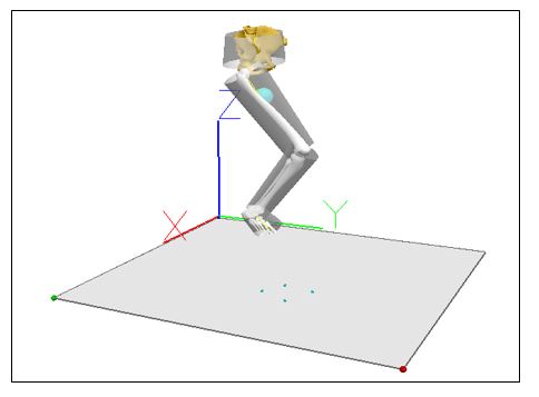
With respect to motion analysis equipment, seven infrared cameras (Oqus 300, Qualisys, Sweden) were set up at a sampling rate of 200 Hz. One force platform (BP12001200, AMTI, USA) with a sampling rate of 2,000 Hz was used to measure ground impact, while an accelerometer (DTS accelerometer 400 g, Noraxon, USA) with a sampling rate set of 500 Hz, which would send signals by a wireless transmitter, was attached to the tibia for investigation of impact mechanism. The direc- tions of the three-dimensional (3-D) spatial coordinates and GRF were established as left (-) and right (+) for the X axis, front (+) and back (-) for the Y axis, and vertical (+) for the Z axis (Figure 1). The Qualisys Track Manager (QTM) program (Qualisys, Sweden) were used to acquire the position data from the reflective markers attached to each joint point and ground reaction force (GRF), while Matlab R2014a (MathWorks, USA) and Visual3D (C-Motion, USA) were used for data analyses. To elim- inate the noise generated while acquiring motion data, smoothing was performed by using a Butterworth second-order low-pass filter, cutoff frequencies for the 3-D coordinate and force platform were set to 12 Hz (Ford, Myer & Hewett, 2007) and 100 Hz (Sell et al., 2007), respectively.
3. Data processing
In the present study, none of the participants performed high-intensity physical activities that can cause fatigue prior to the start of the experiment. The experiment was conducted after receiving a written consent from participants who fully understood the objective and procedures of the study prior to the experiment.
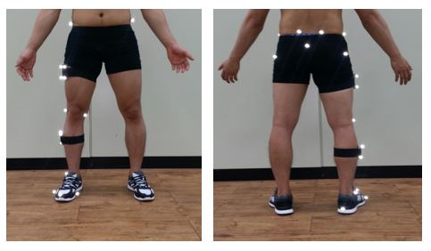
First, the participants' physical characteristics (age, height, and weight) was collected, and the participants performed 10 min of warm-up exercise for safety during the experimental procedures. Then, the par- ticipants wore tights bottom and wore their own shoes. After which, 15 spherical reflective markers on the right lower extremity joints and two marker clusters, one marker each on the thigh and lower leg, were attached to the participants (Figure 2). In addition, an accelerometer was attached to the tibia for investigation of the impact mechanism.
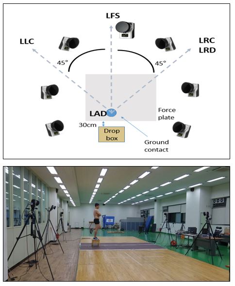
To control the height, a drop box (50 × 40 × 30 cm) was set up 30 cm in an anterior direction from the force platform. Prior to data collection the participants were given prior information on the direction of movement to allow them to perceive the direction of movement in advance, with the direction of movement selected randomly.
To prevent any impact force from being applied during landing, the participants were instructed to perform a single-leg landing by shifting the center of gravity with the left leg extended, only moving the right leg forward. To enable the participants to perform the motion without any difficulties, they were allowed to practice enough trials before per- forming the experimental motion. The motion was performed with both arms crossed and both hands placed under the armpits. Any of the following cases during the experimental motion was considered a failed motion: the foot touched down outside the force platform during landing; mid- or rear-foot landing occurred; the arms moved; and the participant fell from not being able to maintain balance during landing. To analyze the most natural motion, motion was performed 10 trials in each direction (Figure 3).
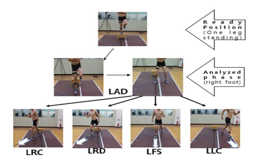
The directions of movement after single-leg landing were defined according to 5 types as follows: landing in place (landing [LAD]), moving the left leg in the right-hand direction by 45° (after landing, right 45° cutting [LRC]), moving the right leg in the right-hand direction by 45° (after landing, right 45° direct [LRD]), moving forward after landing (after landing, forward step [LFS]), and moving the right leg in the left-hand direction by 45° (after landing, left 45° cutting [LLC]). These motions were executed as a continuous motion (Figure 4). For event analysis intervals, events 1, 2, and 3 were set to the moment of touching down on the ground, the moment of peak vertical GRF, and the maximum right-knee flexion angle, respectively.
4. Statistical analysis
For the statistical analysis in the present study, one-way repeated-measures analysis of variance was performed on each variable by using SPSS 21.0 (IBM, USA), with the significance level set at α = .05. If significant effect differences appeared, a post hoc test was performed using Bonferroni corrections.
1. Peak GRF and Acceleration
The analysis of peak vertical GRF and peak vertical acceleration of the lower extremity joints according to the direction of movement showed that the highest peak vertical GRF values (4.17 ± 0.99 and 4.10 ± 0.53 %BW, respectively) were found in movements in the LAD and LLC directions (F = 9.363, p = .000). Moreover, the highest peak vertical acceleration values (26.93 ± 9.34 and 26.35 ± 8.27 g, respec- tively) were found in movements in the LAD and LLC directions. Although significant differences were found in peak vertical GRF (F = 9.363 p = .000), significant differences in peak vertical acceleration were not found (Table 1).
|
|
LAD Mean (SD) |
LRC Mean (SD) |
LRD Mean (SD) |
LFS Mean (SD) |
LLC Mean (SD) |
F (p) |
Post hoc |
ES (ɳ2) |
Power |
|
GRF
peak (%BW) |
|||||||||
|
Peak
vertical |
4.17 (0.99) |
3.35 (1.11) |
3.37 (0.99) |
3.98 (0.61) |
4.10 (0.53) |
9.363 (.000)* |
LRD < LAD LRD < LLC |
.48 |
.99 |
|
Acceleration (g) |
|||||||||
|
Peak
vertical |
26.93 (9.34) |
23.29 (9.82) |
24.16 (10.31) |
23.75 (8.19) |
26.35 (8.27) |
2.113 (.143) |
|
.17 |
.40 |
2. Peak joint power
The analysis of peak joint power of the lower extremity joints according to the direction of movement showed that in the ankle joint, the lowest and highest minimum values (-34.83 ± 11.69 and 23.02 ± 10.39 W/kg, respectively) were found in movements in the LLC and LFS directions (F = 1.179, p = .335). In the knee joint, the lowest and highest minimum values (-18.39 ± 14.39 and -6.97 ± 16.93 W/kg, respectively) were found in movements in the LAD and LFS directions (F = 1.373, p = .261). In both joints, significant differences were not found (Table 2).
|
Peak joint power |
LAD Mean (SD) |
LRC Mean (SD) |
LRD Mean (SD) |
LFS Mean (SD) |
LLC Mean (SD) |
F (p) |
Post hoc |
ES (ɳ2) |
Power |
|
[Sagittal plane] |
|||||||||
|
Ankle |
-32.03 (10.68) |
-26.01 (10.00) |
-23.33 (10.78) |
-23.02 (10.39) |
-34.83 (11.69) |
1.179 (.335) |
|
.10 |
.34 |
|
Knee |
-18.39 (14.30) |
-10.27 (14.48) |
-9.17 (15.19) |
-6.97 (16.93) |
-15.59 (17.39) |
1.373 (.261) |
|
.29 |
.39 |
3. Peak joint moment
The analysis of peak joint moment in the lower extremity joints according to the direction of movement showed that the largest and smallest ankle inversion moments during landing (1.47 ± .64 and 0.99 ± 0.64 Nm/kg, respectively) were found in movements in the LLC and LFS directions (F = 4.298, p = .006). With respect to knee valgus moment, movements in the LAD and LLC directions showed the highest and lowest values of -2.05 ± 1.02 and -1.61 ± 1.05 Nm/ kg, respectively (F = 5.700, p = .006). Moreover, with respect to ankle abduction moment, movements in the LLC and LRC directions showed the highest and lowest values of -0.80 ± 0.27 and -0.58 ± 0.30 Nm/ kg, respectively (F = .600, p = .665). For knee external rotation moment, movements in the LLC and LRC directions showed the highest and lowest values of 0.71 ± 0.27 and -0.59 ± 0.30 Nm/kg, respectively (F = 4.980, p = .014). In the transverse plane, significant differences were found in all the variables, except the ankle joint (Table 3).
|
Peak |
LAD Mean (SD) |
LRC Mean (SD) |
LRD Mean (SD) |
LFS Mean (SD) |
LLC Mean (SD) |
F (p) |
Post hoc |
ES (ɳ2) |
Power |
|
[Frontal
plane] |
|||||||||
|
Ankle (inversion) |
1.23 (0.61) |
1.15 (0.60) |
1.19 (0.55) |
.99 (0.64) |
1.47 (0.64) |
4.298 (.006)* |
LRC < LLC LRD < LLC LFS < LLC |
.30 |
.88 |
|
Knee (valgus) |
-2.05 (1.02) |
-1.96 (1.04) |
-1.82 (1.03) |
-1.79 (1.15) |
-1.61 (1.05) |
5.700 (.006)* |
LLC < LAD LLC < LRC LLC < LFS |
.36 |
.88 |
|
[Transverse plane] |
|||||||||
|
Ankle (abduction) |
-0.66 (.28) |
-0.58 (0.30) |
-0.66 (.33) |
-0.70 (0.34) |
-0.80 (0.27) |
.600 (.665) |
LRC < LFS |
.06 |
.18 |
|
Knee (external rotation) |
-0.68 (0.28) |
-.59 (0.30) |
-0.67 (0.33) |
-.69 (0.37) |
-0.71 (0.27) |
4.980 (.014)* |
LRC < LFS. |
.33 |
.781 |
4. Joint ROM
The analysis of ROM of the lower extremity joints according to the direction of movement showed that in the ankle ROM in the sagittal plane during landing, the highest and lowest ROM (56.59° ± 6.75° and 49.77° ± 7.16°, respectively) were found in movements in the LFS and LAD directions (F = 3.145, p = .024). In the knee joint, the highest and lowest flexion-extension ROM (75.63° ± 18.74° and 50.36° ± 5.67°, respectively) were found in movements in the LRD and LLC directions (F = 14.183, p = .000). In the transverse plane, the highest and lowest ankle ROM (22.06° ± 10.82° and 14.31° ± 5.95°, respectively) were found in movements in the LRC and LLC directions (F = 6.276, p = .068). The highest and lowest knee ROM (25.07° ± 3.00° and 21.87° ± 3.82°, respectively) were found in movements in the LRC direction (F = 1.675, p = .175). In the transverse plane, no significant differences were found in all the variables, except the knee joint (Table 4).
|
Joint ROM |
LAD Mean (SD) |
LRC Mean (SD) |
LRD Mean (SD) |
LFS Mean (SD) |
LLC Mean (SD) |
F (p) |
Post hoc |
ES (ɳ2) |
Power |
|
[Sagittal plane] |
|||||||||
|
Ankle (dorsiflexion) |
49.77 (7.16) |
50.12 (14.13) |
56.51 (6.74) |
56.59 (6.75) |
51.13 (6.38) |
3.145 (.024)* |
LAD < LRC, LAD < LRD LRD < LFS, LAD< LFS LLC < LFS |
.24 |
.77 |
|
Knee (flexion) |
62.05 (11.84) |
71.59 (15.49) |
75.63 (18.74) |
59.76 (16.49) |
50.36 (5.67) |
14.183 (.000)* |
LAD < LRD, LLC < LAD LLC < LRC, LFS < LRD |
.59 |
1.00 |
|
[Transverse plane] |
|||||||||
|
Ankle (adduction) |
18.29 (8.79) |
22.06 (10.82) |
20.45 (12.36) |
19.63 (8.45) |
14.31 (5.95) |
6.276 (.068) |
|
.39 |
.98 |
|
Knee (internal rotation) |
23.08 (5.12) |
25.07 (3.00) |
23.47 (4.46) |
22.47 (5.85) |
21.87 (3.82) |
1.675 (.175) |
LLC < LRC |
.14 |
.47 |
The present study analyzed the kinematic characteristics related to injury factors in the lower extremity joints by using peak vertical GRF, peak vertical acceleration, peak joint power, peak joint moment, and ROM of the lower extremity joints to investigate the injury factors in the lower extremity joints according to the change in direction after single-leg landing.
First, with respect to the impact power at the point of the foot touching the ground, investigation of the peak vertical GRF value for each change in direction during single-leg drop revealed that LAD and LLC directions showed the highest values, with 4.17 ± 0.99 and 4.10 ± 0.53 %BW, respectively, with LAD and LLC showing significant differ- ences. In a study by Cowley, Ford, Myer, Kernozek, and Hewett (2006), these peak GRF values were similar during unexpected cutting motion by basketball and soccer players. Moreover, a study by Kellis and Kouvelioti (2009) showed a peak GRF value during single-leg landing of 4.19 ± 0.40 %BW, which was similar to those in movements in the LAD and LLC directions in the present study. Meanwhile, peak vertical acceleration values were higher in movements in the LAD (26.93 ± 9.34 g) and LLC directions (26.35 ± 8.27 g) than in other directions, which were similar to the peak GRF and peak acceleration values during landing in a study by Tran, Netto, Aisbett and Gastin (2010). Yeh et al. (2013) reported that large impact during landing increased the risk of ankle sprain or knee ACL injury. Thus, large impact being delivered to the lower extremities during landing was found in LAD and LLC, which may cause ligament and joint injuries by transfer to the body.
Investigation of peak joint power in the lower extremity joints based on change in direction after landing showed values of -32.03 ± 10.68 and -18.39 ± 14.30 W/kg for the ankle and knee in LAD, respectively. Moreover, in LLC, the values were -34.83 ± 11.69 and -15.59 ± 17.39 W/kg for the ankle and knee, respectively. In both the ankle and knee joints, significant differences were not found. A study by Kim and Cho (2012) on impact absorption by body segments according to changes in height during drop landing showed similar results, while reporting that increased negative joint power can be viewed as impact absorption capability (Kwon, 2012; Zhang, Bates & Dufek, 2000). Increased negative joint power in the present study predicted the magnitude of impact force in the ankle and knee joints. Impact load is transferred from the ankles to the knees, and increased negative knee joint power is be- lieved to act as a protective mechanism for lowering the risk of ACL injury by absorbing the energy from impact to the knees through eccentric contraction.
With respect to peak moment in the lower extremity joints, ankle inversion moment in LLC showed the highest value of 1.47 ± 0.64 Nm/kg and knee valgus moment showed the lowest value of -1.61 ± 1.05 Nm/kg. Moreover, knee valgus moment showed the highest value in LAD, which was similar to the knee valgus moment reported in a study on injuries during single leg drop conducted by Shimokochi, Ambegaonkar, Meyer, Lee and Shultz (2013). Relatively higher values were seen in the ankle abduction moment (-0.80 ± 0.27 Nm/kg) and knee external rotation moment (-0.71 ± 0.27 Nm/kg) in LLC than in the other conditions. Furthermore, more-significant differences in joint moments were found in the knee joint than in the ankle joint. With respect to knee moments, internal rotation moment has been reported to possibly act as a cause of injury (Meyer & Haut, 2008), but the combined load of exerting external rotation while valgus moment is in effect may also increase the risk of injury (Shimokochi & Shultz, 2008; Shin, Chaudhari & Andriacchi, 2011). Moreover, Kirkendall and Garrett (2000) reported that injuries can be prevented by understanding the role of the inversion moment in the ankles and valgus, and the internal moment and internal rotation in the knee. In the present study, ankle inversion and knee external rotation moments in LLC movement showed a higher risk of anterior talofibular ligament (ATFL) injury than movement in other directions. As LAD had the highest valgus moment, it represented the biggest risk of knee ACL injury.
In the comparison of changes in direction during landing, ankle flexion extension ROM showed the lowest value of 49.77° ± 7.16° in LAD (F = 3.145, p = .024), which was similar to the ankle ROM found in landing with weighted load in a study conducted by Nordin and Dufek (2016) and to 46.56° ± 6.08° found in a study by Kim and Cho (2012) on ankle ROM during single-leg landing according to cutting directions. Moreover, a study by Myer et al. (2015) that compared be- tween single- and double-leg landings also showed a similar pattern of ROM during single-leg landing. Among ankle ROM in executing various movements, all other movements showed higher ankle ROM than LAD, which was because LAD motion involved standing in place without changing directions after landing while perceiving the direction of movement. Thus, impact from GRF, one of the injury mechanisms in the lower extremities, may have been absorbed by ankle flexion extension. In knee flexion extension ROM, the highest and lowest values were found in LRD (75.63° ± 18.74°) and LLC directions (50.36° ± 5.67°), respectively. According to McNair, Marshall and Matheson (1990), ACL injuries occur more often when the knee is extended by about 20° than when it is fully extended. Yeow, Lee and Goh (2009) reported that a large flexion motion is made in the knees to absorb the impact in each joint. In the present study, the highest value was seen in LRD, representing a large impact absorption, which may have appeared so high for absorbing impact delivered to the body. There- fore, ROM showed a high value to reflect rapid transition to the per- ceived direction.
With respect to the injury mechanism in the lower extremity joints, ATFL injuries occurred frequently from repeated landing under the con- dition of ankle plantar flexion and inversion (Safran, Benedettl, Bartolozzi & Mandelbaum, 1999). In the knees, ACL injuries often occurred from hyperextension and valgus of the knees while the feet were firmly planted on the ground, and posterior translational motion and axial rotation of the femur while the tibia was fixed (Neumann, 2010).
In the present study, the peak GRF and peak vertical accelerometer values during landing appeared in LAD and LLC, respectively, which is expected to receive the most impact. Owing to peak joint power, LAD and LLC absorbed more of the impact load on the ankle and knee during landing than LRC and LRD. In the comparison of ROM, LLC showed a lower value than LRC, while LLC showed a lower peak joint moment, which was similar to a study that reported that with respect to peak moment, decrease in knee flexion angle also decreased knee valgus (Lepers, Hausswirth, Mailetti, Briswalter & Hoecke, 2000). There- fore, when changing direction during landing after perceiving the direc- tion of movement, moving to the left is more prone to injury risk than moving to the right. Moreover, LAD movement, which involves no movement after landing while perceiving the direction of movement, poses greater injury risk than other movements.
The present study analyzed biomechanical characteristics of lower extremity joints according to changes in direction during single-leg landing in 11 male participants in their twenties. The study also investi- gated injury mechanism with peak vertical GRF value, peak vertical accelerometer value, peak joint power, joint moment, and ROM.
The highest peak vertical GRF and peak vertical acceleration values were found in LAD. In joint power, the results showed that impact absorption was highest in LAD and LLC, but the differences were not statistically significant. Ankle joint inversion moment was high in LAD and LLC, while knee valgus moment was high in LLC. The biggest difference in ROM was found between LRD and LLC. It is suspected that moving to the left after landing on the right leg is more prone to injury than moving to the right. Moreover, LAD also receives a significant amount of impact, making it prone to injury risk.
Future studies on muscle movement according to the direction of movement during single-leg landing should use the analysis of electro- myography (EMG) and muscle strength in order to understand contri- bution by lower extremity muscles on impact absorption, and studies on non-dominant leg are also warranted.
References
1. Anandacoomarasamy, A. & Barnsley, L. (2005). Long term outcomes of inversion ankle injuries. British Journal of Sports Medicine, 39(3), 1-4.
Crossref
Google Scholar
PubMed
2. Boden, B. P., Dean, G. S., Feagin, J. A. & Garrett, W. E. (2000). Mechanisms of anterior cruciate ligament injury. Orthopedics, 23(6), 573-578.
Crossref
Google Scholar
PubMed
3. Chae, W, S. & Kang, N, J. (2009). Effects of wearing spandex pants on pmpact forces and muscle activities during Drop Landing, Korean Joumal of Sport Biomechanics, 19(3), 603-610.
Crossref
Google Scholar
4. Cho, J, H. Kim, K, H., Moon, G, S. Cho, Y, J. & Lee, S, C. (2010). Analysis of injury mechanism on ankle and knee during drop landings according to landing directions, Korean Joumal of Sport Biomech- anics, 20(1), 67-73.
Crossref
Google Scholar
5. Choi, J. K. (2015). Kinetic comparison analysis about normal landing and intentional landing for safety at drop landing movement. The Koeran Journal of Sports Science, 23(6), 1467-1477.
Crossref
6. Cowley, H. R., Ford, K. R., Myer, G. D., Kernozek, T. W. & Hewett, T. E. (2006). Differences in neuromuscular strategies between landing and cutting tasks in female basketball and soccer athletes. Journal of Athletic Training, 41(1), 67-73.
Crossref
Google Scholar
7. Ford, K. R., Myer, G. D. & Hewett, T. E. (2007). Reliability of landing 3D motion analysis: implications for longitudinal analyses. Medicine and Science in Sports and Exercise, 39(11), 20-21.
Crossref
Google Scholar
PubMed
8. Gehring, D., Melnyk, M. & Gollhofer, A. (2009). Gender and fatigue have influence on knee joint control strategies during landing. Clinical Biomechanics, 24(1), 82-87.
Crossref
Google Scholar
PubMed
9. Hang, B. (2013). Acute sports-related lower extremity injuries. Clinical Pediatric Emergency Medicine, 14(4), 304-317.
Crossref
Google Scholar
10. Hewett, T. E., Myer, G. D. & Ford, K. R. (2004). Decrease in neuromuscular control about the knee with maturation in female athletes. The Journal of Bone & Joint Surgery, 86(8), 1601-1608.
Crossref
Google Scholar
PubMed
11. Hootman, J. M., Dick, R. & Agel, J. (2007). Epidemiology of collegiate injuries for 15 sports: summary and recommendations for injury prevention initiatives. Journal of Athletic Training, 42(2), 311-319.
Crossref
Google Scholar
PubMed
12. Houck, J. R., Duncan, A. & De Haven, K. E. (2006). Comparison of frontal plane trunk kinematics and hip and knee moments during anticipated and unanticipated walking and side step cutting tasks. Gait & Posture, 24(3), 314-322.
Crossref
Google Scholar
13. Houck, J. R., Wilding, G. E., Gupta, R., De Haven, K. E. & Maloney, M. (2007). Analysis of EMG patterns of control subjects and subjects with ACL deficiency during an unanticipated walking cut task. Gait & Posture, 25(3), 628-638.
Crossref
Google Scholar
PubMed
14. Kellis, E. & Kouvelioti V. (2009). Agonist versus antagonist muscle fatigue effects on thigh muscle activity and vertical ground reaction during drop landing. Journal of Electromyography and Kinesiology, 19(1), 55-64.
Crossref
Google Scholar
15. Kim, K. H. & Cho. J. H. (2012). The influence of cutting direction on risk factors of anterior curuciate ligamentlijury. Journal of Sport and Leisure Studies, 48(2), 795-802.
Crossref
16. Kim, J. S., Oh, J. H. & Jeong, I. S. (2015). A biomechanical analysis on the action of volleyball vertical jumping performance. Journal of Korean Society for the Study of Physical Education, 19(4), 131-143.
Crossref
17. Kirkendall, D. T. & Garrett, W. E. (2000). The anterior cruciate ligament enigma: injury mechanisms and prevention. Clinical Orthopaedics and Related Research, 372, 64-68.
Crossref
Google Scholar
18. Kwon, M. S. (2012). Effect of added mass between male and female on the lower extremity joints angular velocity, moment, Absorb energy during drop landing. Korean Joumal of Sport Biomechanics, 22(3), 325-332.
Crossref
19. Kwon, O. B., Jung, C. J., Park, K. J., Kwon, M. S. & Shin, S. H. (2007). The analysis of joint motion and moment of Lower extremities to cutting angles. Korean Journal of Physical Eduaction, 46(2), 451-459.
Crossref
Google Scholar
20. Lepers, R., Hausswirth, C., Maffiuletti, N., Brisswalter, J. & van Hoecke, J. (2000). Evidence of neuromuscular fatigue after prolonged cycling exercise. Medicine and Science in Sports and Exercise, 32(11), 1880 -1886.
Crossref
Google Scholar
PubMed
21. Lee, K. K. (1998). The effect of turning direction on lower extremity joint moment. Journal of Biomechanics, 8(2), 21-42.
Crossref
Google Scholar
22. Majewski, M., Susanne, H. & Klaus, S. (2006). Epidemiology of athletic knee injuries: A 10-year study. The Knee, 13, 184-188.
Crossref
Google Scholar
PubMed
23. McNair, P., Marshall, R. & Matheson, J. (1990). Important features asso- ciated with acute anterior cruciate ligament injury. The New Zealand Medical Journal, 103(901), 537-539.
Crossref
Google Scholar
24. McNitt-Gray, J. L. (1989). The influence of impact speed on joint kine- matics and impulse characteristics of drop landings. Journal of Biomechanics, 22(10), 1054.
Crossref
25. Meyer, E. G. & Haut, R. C. (2008). Anterior cruciate ligament injury induced by internal tibial torsion or tibiofemoral compression. Journal of Biomechanics, 41(16), 3377-3383.
Crossref
Google Scholar
26. Myer, G. D., Bates, N. A., Dicesare, C. A., Foss, K. D. B., Thomas, S. M., Wordeman, S. C., Suqimoto, D., Roewer, B. D., Mckeon, J. M. M., Stasi, S. L. D., Noehren, B. W., McNally, M., Ford, K. R., Kiefer, A. W. & Di Stasi, S. L. (2015). Reliability of 3-dimensional measures of single-leg drop landing across 3 institutions: implications for multi- center research for secondary ACL-injury prevention. Journal of Sport Rehabilitation, 24(2), 198-209.
Crossref
27. Neumann D. A. (2010). Kinesiology of the musculoskletal system: foun- dations of rehabilitation 2nd ed, St. Louis, MO: Mosby/Elsevier
Crossref
28. Nordin, A. D. & Dufek, J. S. (2016). Neuromechanical synergies in single-leg landing reveal changes in movement control. Human Movement Science, 49, 66-78.
Crossref
Google Scholar
29. Safran, M. R., Benedetti, R. S., Bartolozzi 3rd, A. & Mandelbaum, B. R. (1999). Lateral ankle sprains: a comprehensive review: part 1: etio- logy, pathoanatomy, histopathogenesis, and diagnosis. Medicine and Science in Sports and Exercise, 31(7), 429-437.
Crossref
Google Scholar
30. Schmitz, R. J., Kulas, A. S., Perrin, D. H., Riemann, B. L. & Shultz, S. J. (2007). Sex differences in lower extremity biomechanics during single leg landings. Clinical Biomechanics, 22(6), 681-688.
Crossref
Google Scholar
31. Sell, T. C., Ferris, C. M., Abt, J. P., Tsai, Y. S., Myers, J. B., Fu, F. H. & Lephart, S. M. (2007). Predictors of proximal tibia anterior shear force during a vertical stop-jump. Journal of Orthopaedic Research, 25(12), 1589-1597.
Crossref
Google Scholar
32. Shin, E.S., Choi, J. K. & Kim, S. D. (2015). Kinematical analysis of the causes of injury during landing in floor exercises by middle school male athletes. The Korean Journal of Sports Science, 24(3), 1693-1702.
Crossref
33. Shimokochi, Y., Ambegaonkar, J. P., Meyer, E. G., Lee, S. Y. & Shultz, S. J. (2013). Changing sagittal plane body position during single-leg landings influences the risk of non-contact anterior cruciate liga- ment injury. Knee Surgery, Sports Traumatology, Arthroscopy, 21(4), 888-897.
Crossref
Google Scholar
34. Shimokochi, Y. & Shultz, S. J. (2008). Mechanisms of noncontact anterior cruciate ligament injury. Journal of Athletic Training, 43(4), 396-408.
Crossref
Google Scholar
PubMed
35. Shin, C. S., Chaudhari, A. M. & Andriacchi, T. P. (2011). Valgus plus internal rotation moments increase anterior cruciate ligament strain more than either alone. Medicine and Science in Sports and Ex- ercise, 43(8), 1484-1491.
Crossref
Google Scholar
36. Sigward, S., Pollard, C. & Powers, C. (2012). The influence of sex and maturation on landing biomechanics: implications for anterior cruciate ligament injury. Scandinavian Journal of Medicine & Science in Sports, 22(4), 502-509.
Crossref
Google Scholar
37. Taylor, J. B., Ford, K. R., Nguyen, A. D. & Shultz, S. J. (2016). Bio- mechanical comparison of single- and double-leg jump landings in the sagittal and frontal plane. American Orthopaedic Society for Sports Medicine, 4(6) 1-9.
Crossref
Google Scholar
38. Tran, J., Netto, K., Aisbett, B. & Gastin, P. (2010). Validation of accel- erometer data for measuring impacts during jumping and landing tasks. International Society of Biomechanics in Sports, 1-4.
Crossref
Google Scholar
39. Tropp, H., Askling, C. & Gillquist, J. (1985). Prevention of ankle sprains. The American Journal of Sports Medicine, 13(4), 259-262.
Crossref
Google Scholar
PubMed
40. Yeh, C. C., Chang, S. F., Huang, T. Y., Chang, H. I., Kuo, H. C., Wu, Y. C. & Chen, C. N. (2013). Shear stress modulates macrophage-induced urokinase plasminogen activator expression in human chondrocytes. Arthritis Research & Therapy, 15(2), 1-13.
Crossref
Google Scholar
41. Yeow, C., Lee, P. & Goh, J. (2009). Effect of landing height on frontal plane kinematics, kinetics and energy dissipation at lower extre- mity joints. Journal of Biomechanics, 42(12), 1967-1973.
Crossref
Google Scholar
PubMed
42. Yeow, C. H., Lee, P. V. S. & Goh, J. C. H. (2011). An investigation of lower extremity energy dissipation strategies during single-leg and double-leg landing based on sagittal and frontal plane biomech- anics. Human Movement Science, 30(3), 624-635.
Crossref
Google Scholar
43. Zhang, S. N., Bates, B. T. & Dufek, J. S. (2000). Contributions of lower extremity joints to energy dissipation during landings. Medicine and Science in Sports and Exercise, 32(4), 812-819.
Crossref
Google Scholar
