Open Access, Peer-reviewed
eISSN 2093-9752
Open Access, Peer-reviewed
eISSN 2093-9752
Ji-Seon Ryu
http://dx.doi.org/10.5103/KJSB.2016.26.1.31 Epub 2016 April 20
Abstract
The purpose of this study was to investigate the differences in FT (free-torque) components between non-fatigue and fatigue conditions induced by prolonged running.
Fifteen healthy runners with no previous lower-extremity fractures (22.0 ± 2.1 years of age) participated in this study. Ground reaction force data were collected for the right-stance phase for 10 strides of 5 and 125-min running periods at 1,000 Hz using an instrumented force platform (instrumented dual-belt treadmills, Bertec, USA) while the subjects ran on it. The running speed was set according to the preferences of the subjects, which were determined before the experiment. FT variables were calculated from the components of the moment and force output from the force platform. A repeated- measures one-way ANOVA was used to test for significant differences between the two conditions. The alpha level for all the statistical tests was 0.05.
The absolute FT at the peak braking force was significantly greater after 5 mins of running than after 125 mins of running—which was regarded as a fatigued state— but there were no significant differences in the absolute peak FT or impulse between the conditions.
The FT variables in the fatigue condition during prolonged running hardly affect the tibial stress syndrome.
Keywords
Free torque Impulse Fatigue Prolonged running Tibial stress syndrome Ground reaction force
Running is a popular sport because it demands physical activity and can be performed at a convenient time in any place (Ida et al., 2010). Although running has a positive effect on health, it can have a negative impact on the body because of possible injury. In particular, the incidence of injury from prolonged running is reported to be 30~ 79% (Lun et al., 2004; Taunton et al., 2003; van Gent et al., 2007). There are various possible factors involved in injury from prolonged running, including mechanical abnor- malities previous injuries sex, body mass index, training frequency, intensity, and duration, muscle strength, flexibility, shoes, and fatigue (Taunton et al., 2003).
Among prolonged running injuries, stress fractures are a common problem and account for a large proportion of running injuries (Taunton et al., 2002). In prolonged running, stress fractures of the tibia are most common, accounting for 35~56% of the stress fractures caused by prolonged running (Romani et al., 2002). One of the bio- mechanical factors causing tibial stress fractures is free torque (FT) (Milner & Davis, 2006). FT refers to the torque in the vertical axis that is generated by friction between the foot and the ground during the support phase in running (Holden & Cavanagh, 1991). Because the tibia is positioned proximal to the foot, FT is transferred to the tibia as a twisting load, which is responsible for stress fractures of the tibia during running (Pohl et al., 2008). Holden & Cavanagh (1991) reported that FT is closely related to the pronation of the foot. In a retrospective study, Milner et al. (2006) discovered that experienced runners with a past tibial stress fracture developed the in- jury because they had larger absolute FT than runners who had no tibial stress fractures. In another retrospective study, Pohl et al. (2008) claimed that the FT during running was the most important variable of interest for predicting tibial stress fractures in female runners. Other studies have re-ported that the abnormal absolute FT during running causes stress fractures by altering the normal load patterns on the tibia. Although studies report that runners who naturally generate large a FT during running have a larger risk of tibial stress fractures, very few studies have investi- gated whether the effect of FT on tibial stress fractures depends on the running time or whether increased fatigue with longer running times worsens the effect of FT on tibial stress fractures. There are no quantitative results available on whether runners with a naturally high FT have a higher risk of tibial stress fractures or whether acquired factors such as fatigue can cause stress fractures by increasing the FT. Among the motor factors that can affect the FT, foot pronation (Holden & Cavanagh, 1991) and the medial rotation of the tibia have been reported to increase in states of fatigue (Ryu, 2001). Thus, it is considered that FT levels should change in a fatigued state, but concrete observations are required. Because fatigue-related injuries usually occur in the latter stages of exercise (Collins & Whittle, 1989), investigating the FT, which is a cause of tibial stress fractures, under conditions of fatigue is an important task that can provide key insights from a clinical perspective and regarding exercise performance. Thus, the FT in pro- longed running, which influences tibial stress injuries, must be analyzed in conditions of fatigue, according to the running duration.
Fatigue refers to the state in which muscles no longer respond to the normal level of contraction signals (Kang et al., 2008; Ryu, 2001, 2013), making it impossible to main- tain a certain level of tension (Asmussen, 1979). Fatigue is accompanied by muscular discomfort and pain, and when exercise exceeds aerobic energy production levels or energy sources are depleted owing to insufficient recovery time, there is an accumulation of metabolic byproducts such as lactate (Korean Society of Exercise Physiology [KSEP], 2014). Because fatigue impairs muscle strength, coordination, mental concentration, and attention, it increases the risk of injury (Collins & Whittle, 1989). In prolonged running, although fatigue increases with time and distance, it is important to recognize that the precise level of fatigue differs according to the composition of muscle-fiber types, exercise intensity, contraction time, and individual stamina. Nevertheless, at 70% maximal oxygen uptake (vo2 max) or 75~80% of the maximal heart rate, a sudden feeling of fatigue is reported to occur after 1 hr (Ryu, 2001, 2013; Wilmore & Costill, 1994). This means that when running, to achieve a state of complete muscle fatigue, an individual must run at the aforementioned exercise intensity for at least 1 hr, and when running at a lower intensity, the individual must run for longer (Ryu, 2013).
Several studies have been performed to evaluate bio- mechanical abnormalities that cause injury in a state of prolonged running-induced fatigue, investigate the relation- ship between exercise and biomechanical factors in order to predict the potential occurrence of injury, and diag- nose and understand injury (Bruggemann & Arndt, 1994; Bruggemann et al., 1995; Dierks et al., 2010; Gheluwe et al., 1995; Hunter & Smith, 2000; Jean-Benoit et al., 2011; Nicol et al., 1991; Paavolainen et al., 1995; Ryu, 2013; Ryu, 2014; Siler & Martin, 1991; Verbitsky et al., 1998; Williams et al., 1991). However, no studies have investigated the FT—which is known to be a cause of tibial stress fractures during running—in a state of fatigue.
Therefore, we aimed to observe and compare the abso- lute FT in the early stages (5 mins) of running and in a state of fatigue after 125 mins of running. To this end, we focused on the FT amplitude at the passive force peak within the support phase, the impulse representing the area of the FT multiplied by time during the support phase, and the maximal absolute FT during the support phase. Differences between the variables analyzed in this study at different time points were assumed to be the result of fatigue.
1. Subjects
We selected 15 healthy subjects in their 20s who were able to run for at least 2 hrs at their preferred speed and had a rearfoot strike. Their physical characteristics and preferred running speeds are shown in Table 1.
|
Age |
Height |
Mass |
Preferred run speed (m/s) |
|
22.0 ± 2.1 |
175.8 ± 4.3 |
66.1 ± 5.1 |
2.5 ± 0.1 |
2. Experimental procedure
All the subjects were made to run at their preferred speed on an instrumented dual-belt treadmill (Bertec, USA), and measurements of six components of the ground reaction force (GRF; Fx, Fy, Fz, Mx, My, Mz) were collected at a sampling rate of 1,000 Hz for 10 strides (20 steps) upon the striking of the right foot. To reduce the variance due to differences in footwear, all subjects wore the same training shoes (Lunareclipse2, Nike, USA) and ran for 2 hrs and 10 mins. Although fatigue is subject to individual differences, previous studies reported that individuals gene- rally feel fatigue after running for 1 hr at 70% of their vo2 max or 75~80% of their maximal heart rate (Ryu, 2001, 2013; Wilmore & Costill, 1994). Therefore, in this study, we defined the experimental conditions for complete fatigue as having run for at least 2 hrs (Ryu, 2013). Data were collected after 5 and 125 mins of running in a manner that could not be perceived by the subjects. Prior to the data collection, all the subjects warmed up sufficiently and were allowed to become familiar with running on the treadmill
3. Data processing and analysis
The coordinates for this study were defined according to the right-hand rule: the forward direction for the runner was +y, the upward direction was +z, and the right-hand side of the cross between these two directions was +x. The absolute FT value was used at the moment when the foot contacted the ground, irrespective of pronation or supination. Before calculating the FT, the cutoff frequency was calculated to filter the signal for all GRF data, con- sisting of three directional-force and three axial-moment components. The cutoff frequency was determined accord- ing to the signal power spectrum density (PSD) as the peak frequency, which was defined as the frequency at a cumu- lative power of 99.9% (Ryu, 2013; Stergiou et al., 2002). Next, to remove the direct-current component from the signal, the mean of the first five points from the signal was calculated and subtracted from the whole signal (Ryu, 2013). The range of analysis was limited to the support phase, i.e., from the moment the foot touched the ground to the moment the foot left the ground. Specifically, foot-down and foot-up were defined as vertical GRF ≥ 5 N and < 5 N, respectively, and a rectangular window was applied to the data for this phase (Ryu, 2013). The support phase for the remaining five GRF components was defined according to the vertical GRF component. From the pro- cessed GRF signal, we calculated the FT in the vertical axis due to the friction between the foot and the ground. The FT was calculated using the moment acting in the vertical axis (Mz) obtained from the origin of the GRF and the moment due to the total shear stress acting through the center of pressure (CoP) (Holden & Cavanagh, 1991). That is, the FT was calculated using the following equation describing the roles of these two components representing the vertical moment from the output of the force plate (Milner et al., 2006).
Here, MZ is the moment in the z-axis, CoPX is the x-coordinate of the CoP, FY is the GRF in the y-direction, CoPY is the y-coordinate of the CoP, FZ is the GRF in the z-direction, and MX is the moment in the x-axis. CoPY was adjusted according to the running speed and sampling rate.
For example, given the sampling rate of 1,000 Hz used in this study, at a running speed of 2.5 m/s, multiples of 0.0025 m were added to each sample point.
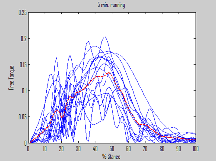
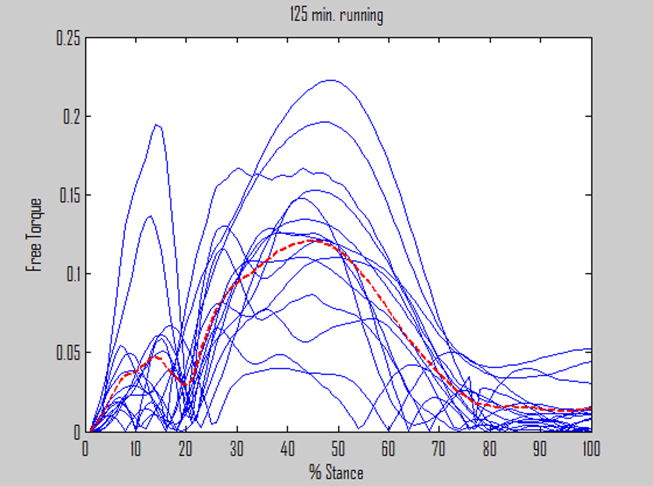
To reduce the impact of physical differences among the subjects on the FT, the FT was normalized according to the height and body weight to make it a dimensionless variable before the FT amplitude at the peak passive force in the support phase; the total impulse—calculated as the area under the FT curve over the whole support phase; and the maximum absolute FT during the support phase were calculated. All the variables were calculated and aver- aged over 5 and 10 strides for all subjects, and the norma- lized FT was plotted.
To evaluate the differences in the calculated variables for different running times-corresponding to fatigue and non-fatigue—a repeated measure one-way ANOVA was performed, and a statistically significant level of α = 0.05 used for all comparisons.
We observed the absolute FT in a state of prolonged running-induced fatigue according to the aforementioned method. Table 2 shows the results of the statistical analysis of the differences between time points. Graphs of the nor- malized FT with respect to time in the support phase of running are shown in Figs. 1 and 2. According to the results, at the peak passive magnitude of the vertical GRF signal during the support phase, the FT (mean ± SD) was 0.067 ± 0.038 after 5 mins of running and 0.037 ± 0.26 after 125 mins of running. The FT after 5 mins was signifi- cantly higher (p < 0.05). The maximum amplitude of the absolute FT during the support phase was 0.153 ± 0.26 after 5 mins, which was slightly larger than 0.140 ± 0.041 after 125 mins, but this difference was not statistically significant. The impulse, measured as the area under the FT × time curve, was 0.017 ± 0.004 after 5 mins of run- ning, which was slightly larger than the value of 0.016 ± 0.004 after 125 mins. However, as in the case of the abso- lute FT, there was no significant difference between the two running-time conditions.
|
Variables Conditions |
Free torque |
Absolute |
Impulse |
|
5 mins 125 mins |
0.067 ± 0.038 0.037 ± 0.026 |
0.153 ± 0.026 0.140 ± 0.041 |
0.017 ± 0.004 0.016 ± 0.004 |
|
F value p values |
5.92 0.02 |
1.32 0.25 |
1.17 0.28 |
We aimed to investigate changes in the FT during run- ning in a state of prolonged running-induced fatigue. The analyzed FT variables were the absolute FT upon braking; the maximum absolute FT during the support phase; and the impulse, representing the product of the FT and time during the support phase. Although the individual differ- ences in the FT at the passive force peak were large, the values at 5 mins, without fatigue, were significantly larger than the values at 125 mins, with fatigue. Nevertheless, the maximum absolute FT and the total impulse in the support phase were larger at 5 mins than 125 mins, but there was no statistically significant difference.
In the support phase of running, the shear stress, moment, and FT are important indicators for predicting injury at the moment of stopping (Milner et al., 2006). For prolonged running, Jean-Benoit et al. (2011) and Ryu (2013) reported that there was no significant difference in the impulse between fatigued and non-fatigued states. Finni et al. (2003) and Ryu (2013) also reported no significant difference in the shear stress acting in the mediolateral or anterio- posterior direction in a state of fatigue caused by increased running time. According to these previous studies, a poten- tial reason for the FT being smaller at 125 mins was the increase in the moment acting in the forward and lateral directions, which caused a large change in the CoP and a decrease in the FT. Another possibility is that a decrease in the impulse peak (Gerlach et al., 2005) resulted in a large CoP and a small overall FT.
To gather evidence on this, future studies that investi- gate the overall relationships between the FT, the CoP, and the moment acting in the forward and mediolateral directions are required. Because exercise variables play an important role in altering the normal alignment of the body during running (Milner et al., 2006) and causing stress fractures, additional studies on the difference between the two conditions in the FT during stopping using observa- tions of the joints and segments of the body and inves- tigations of changes in the FT are required.
As previously mentioned, running has positive effects on physical condition, including endurance and muscle strength, but 19~79% of runners are reported to suffer injuries each year (Hasegawa et al., 2007; van Gent et al., 2007). Risk factors for running injuries are related to the cause of injury. A common injury that can develop from running is a stress fracture, with an annual incidence of 6~14% in runners (Taunton et al., 2002). The tibial crest twists while running because of the FT acting between the foot and the ground. The FT is generated by the move- ments of the runner on the ground in order to regulate the angular momentum of the body in the horizontal plane (Willwacher et al., 2015). This FT is transferred as a twisting load on the tibia and therefore acts as a risk factor that can cause pain and injury (Milner et al., 2006; Willwacher et al., 2015). The tibia is the bone that is most easily exposed to stress fractures during running and accounts for 35~56% of all stress injuries (Romani et al., 2002). According to previous studies, the twisting load—or FT—acting on the tibia is larger in the population who experience tibial stress injuries than in the healthy popu- lation (Milner et al., 2006). Moreover, individuals who have experienced a tibial stress fracture are known to show a recurrence rate of 36% (Hauret et al., 2001). Pohl et al. (2008) claimed that the FT during running was a risk factor for tibial stress, as it accurately predicted a past history of tibial stress fracture in 83% of cases. Thus, the FT is known to act as a mechanical variable that causes fractures by increasing the load on the tibia during running.
Prolonged running and other repetitive activities can easily expose the body to overuse injuries. The potential increase in injuries during prolonged running is reported to occur in the latter stages of running, when fatigue is usually elevated (Whiting & Zerinicke, 1998). That is, with prolonged running, increases in distance and time create fatigue and can thus be considered to yield a high risk of injury (Brill & Macera, 1995). Therefore, observing injury risk factors after prolonged running to induce fatigue is an important task for providing a means of preventing injury by regulating the risk factors in runners vulnerable to injury. Examining the risk factors for injury in a state of fatigue can be considered an attempt to reduce potential injuries during prolonged running (Whiting & Zerinicke, 1998). As previously discussed, there is a lack of research on observing the FT that influences the twisting of the tibia during prolonged running in a state of fatigue.
Although this study did not demonstrate a statistically significant relationship, the maximum absolute FT was approximately 9% lower in the support phase at 125 mins, which is thought to have contributed to a decrease in the active component of the vertical GRF (Christina, White, & Gilchrist, 2001; Nicol et al., 1991; Ryu, 2013). During pro- longed running, injuries are more common under fatigue. This study observed the FT, which causes tibial stress fractures, in a state of fatigue and found that although the FT at the peak passive force decreased in a fatigued state, there was no change in the maximum absolute FT according to the exercise amount. Hence, the negative effect of the FT on tibial stress was not confirmed in a state of short-term fatigue. According to the results of this study, the FT is a congenital issue for individual runners and is not influenced by the acquired factor of fatigue.
We observed the effects of fatigue induced by prolonged running on a treadmill on the components of the FT. We analyzed 15 males in their 20s with a rearfoot strike and no experience of lower-limb injury.
To conduct related studies in the future, we propose the need to analyze the relationship between the CoP and FT in the lower limb in a state of running-induced fatigue. Furthermore, the FT components should be observed in a state of long-term accumulated fatigue rather than only short-term fatigue.
Although the results showed slightly larger values for the maximum absolute FT and impulse in the support phase at 5 mins compared with 125 mins, this difference was not statistically significant. Nevertheless, the absolute FT at the peak passive force was larger at 5 mins than at 125 mins, exhibiting a significant reduction after 2 hrs of running (p < 0.05). These results show that prolonged running-induced fatigue did not have a large effect on the potential for tibial stress fractures via its impact on FT components.
References
1. Asmussen, E. (1979). Muscle fatigue. Medicine and science in sports, 11(4), 313-321.
Crossref
Google Scholar
PubMed
2. Brill, P. A. & Macera, C. A. (1995) The influence of running patterns on running injuries. Sports medicine. 20, 365 -368.
Crossref
Google Scholar
3. Brüggemann, G. P. & Arndt, A. (1994). Fatigue and lower extremity function. In Proceedings of the First Sympo- sium on Functional Footwear. Calgary (pp. 4-5).
Crossref
4. Brüggemann, G. P., Arndt, A., Kersting, U. G. & Knicker, A. J. (1995). Influence of fatigue on impact force and rearfoot motion during running. Proceedings of Inter- national Society of Biomechanics. 132-133.
Crossref
5. Collins, J. & Whittle, M. W. (1989). Impulsive forces during walking and their clinical implications. Clinical Bio- mechanics, 4, 179-187.
Crossref
Google Scholar
6. Christina, K. A., White, S. C. & Gilchrist, L. A. (2001). Effect of localized muscle fatigue on vertical ground reaction forces and ankle joint motion during running. Human movement science. 20, 257-276.
Crossref
Google Scholar
7. Dierks, T. A., Davis, I. S. & Hamill, J. (2010). The effects of running in an exerted state on lower extremity kine- matics and joint timing. Journal of Biomechanics, 43(5), 2993-2998.
Crossref
Google Scholar
PubMed
8. Finni, T., Kyröläinen, Avela, J. & Komi, P. V. (2003). Maximal but not submaximal performance is reduced by constant-speed 10-km run. Journal of Sports Medicine and Physical Fitness, 43(4), 411-417.
Crossref
Google Scholar
9. Gheluwe, B. V., Kopriva, N. & Madsen, C. (1995). Rearfoot motion in running prior to volitional exhaustion. Pro- ceedings of XV Congress of International Society of Biomechanics, 958-959.
Crossref
10. Gerlach, K. E., White, S, C., Burton, H. W., Dorn, J. M., Leddy, J .J. & Horvath, P. J. (2005). Kinetic changes with fatigue and relationship to injury in female runners. Medicine and science in sports and exercise. 37(4), 657-663.
Crossref
Google Scholar
11. Hasegawa, H., Yamauchi, T. & Kraemer, W. J. (2007). Foot strike patterns of runners at the 15km point during an elite-level half marathon. Journal of strength and conditioning Research. 21(3), 888-893.
Crossref
Google Scholar
12. Hauret, K. G., Shippey, D. L. & Knapik, J. J. (2001). The physical training and rehabilitation program: duration of rehabilitation and final outcome of injuries in basic combat training. Military Medicine. 166, 820-826.
Crossref
Google Scholar
13. Hunter, I. & Smith, G. A. (2000). Effect of fatigue on pre- ferred and most economical stride frequency in tread- mill running. Proceedings of XIth Congress of the Canadian Society for Biomechanics, 43.
Crossref
14. Holden, J. P. & Cavanagh, P. R. (1991). The free moment of ground reaction in distance running and its changes with pronation. Journal of Biomechanics. 24, 887-897.
Crossref
Google Scholar
PubMed
15. Ida, B., Steef W. B., Koen A. P. M., Lemmink, Willem van Mechelen, M. D. & Ron L. D. (2010). Predictors of running-related injuries in Novice runners enrolled in a systematic training program, The American Journal of Sports Medicine, 38(2), 273-280.
Crossref
Google Scholar
16. Jean-Benoit, M., Pierre, S. & Guillaume, Y. H. (2011). Changes in running kinematics, kinetics, and spring-mass be- havior over a 24-h run. Medicine & Science in Sports & Exercise, 43(5), 829-836.
Crossref
Google Scholar
PubMed
17. Kang, H. M., Kim, D. H., Seo, J. R., Yi, Y. W. & Park, S. K. (2008). Muscle fatigue compensating exoskeleton. The Korean Society of Mechanical Engineers. 5, 191-192
18. Korean society of exercise physiology. (2014). Exercise physi- ology, Hanmibook, 123.
19. Lun, V., Meeuwisse, W. H., Stergiou, P. & Stefanyshyn, D. (2004). Relation between running injury and static lower limb alignment in recreational runners. British journal of sports medicine. 38, 576-580.
Crossref
Google Scholar
PubMed
20. Milner, C. E., Davis, I. S. & Hamill, J. (2006). Free moment as predictor of tibial stress fracture in distance runners. Journal of Biomechanics. 39, 2819-2825.
Crossref
Google Scholar
PubMed
21. Nicol, C., Komi P. V. & Marconnet, P. (1991). Fatigue effects of marathon running on neuromuscular performance changes in muscle force and stiffness characteristics. Scandinavian. Journal of Medicine & Science in Sports, 10-17.
Crossref
Google Scholar
22. Paavolainen, L., Hakkinen, K., Nummela, A. & Ruskoh, (1995). Effects of fatigue on stride parameters in en- durance athletes with different distance running per- formance capability. Proceedings of XV Congress of International Society of Biomechanics.
23. Pohl, M. B., Mullineaux, D. R., Milner, C. E., Hamill, J. & Davis, I. S. (2008). Biomechanical predictors of retrospective tibial stress fractures in runners. Journal of Biomech- anics, 41, 1160-1165.
Crossref
Google Scholar
PubMed
24. Romani, W. A., Gieck, J. H., Perrin, D. H., Saliba, E. N. & Kahler, D. M. (2002). Mechanisms and management of stress fracures in physically active persons. Jounal of Athletic Training, 37, 306-314.
Crossref
Google Scholar
PubMed
25. Ryu, J. S. (2001). Fatigue effects on biomechanical para- meters during a prolonged run. The Korean Journal of Physical Education, 40(4), 1011-1025.
26. Ryu, J. S. (2013). Effects of a prolonged-run-induced fatigue on the ground reaction force components. Korean Journal of Sport Biomechanics, 23(3), 225-233.
Crossref
Google Scholar
27. Ryu, J. S. (2014). Variability of GRF components between increased running times during prolonged run. Korean Journal of Sport Biomechanics, 24(4), 359-365.
Crossref
Google Scholar
28. Siler, W. L. & Martin, P. E. (1991). Changes in running pat- tern during a treadmill run to volitional exhaustion: Fast vs slow runners. International Journal of Sports Biomechanics, 7, 12-28.
Crossref
29. Stergiou, N., Giakas. G., Byrne, J. E. & Pomeroy. V. (2002). Frequency domain characteristics of ground reaction forces during walking of young and elderly females. Clinical Biomechanics, 17, 615-617.
Crossref
Google Scholar
30. Taunton, J. E., Ryan, M. B., Clement, D. B., McKenzie, D. C., Lloyd-Smith, D. R. & Zumbo, B. D. (2002). A retro- spective case-control analysis of 2002 running injuries. British Journal of Sports Medicine 36, 95-101.
Crossref
Google Scholar
PubMed
31. Taunton, J. E, Ryan, M. B, Clement, D. B, McKenzie, D. C, Lloyd-Smith, D. R. & Zumbo, B. D. (2003). A prospec- tive study of running injuries: the Vancouver Sun Run
Crossref
Google Scholar
PubMed
32. van Gent, R. M., Siem, D., van Middlekoop M., van Os A. G., Bierma-Zeinstra, A. M. A. & Koes B. W. (2007). In- cidence and determinants of lower extremity running injuries in long distance runners: a systematic review. British journal of sports medicine. 41, 469-480
Crossref
Google Scholar
33. Verbitsky, O., Mizrahi, J., Voloshin, A., Treiqer, J. & Elisakov, (1998). Shock transmission and fatigue in human run- ning. Journal of Applied Biomechanics, 14, 301-311.
Crossref
34. Williams, K. R., Snow, R. & Agruss, C. (1991). Changes in distance running kinematics with fatigue. International Journal of Sports Biomechanics, 7, 138-162.
Crossref
Google Scholar
35. Willwacher, S., Fischer, K., Goetze, I., Hamill, J., Rohr, E. & Bruggemann, G. P. (2015). Free moment patterns, trans- versal plane joint loading and injury risk in running. Footwear Science. 7(S1), s124-126.
Crossref
Google Scholar
36. Wilmore, J. H. & Costill, D. L. (1994). Physiology of sport and exercise. Human Kinetics, 110-522.
Crossref
Google Scholar
37. Whiting, W. C. & Zerinicke, R. F. (1998). Biomechanics of Musculoskeletal injury. Champaign, IL. Human Kinetics, 119.
