Open Access, Peer-reviewed
eISSN 2093-9752
Open Access, Peer-reviewed
eISSN 2093-9752
Ji-Seon Ryu
http://dx.doi.org/10.5103/KJSB.2016.26.3.257 Epub 2016 October 15
Abstract
Objective: The purpose of this study was to determine the periodicity of shank-foot segment coupling and free torque before and after fatigue induced by prolonged running.
Method: Fifteen young healthy male participants with a rear-foot strike ran on instrumented dual-belt treadmills at 70% of their maximum oxygen uptake for 65 min. Kinematic and ground reaction force data were collected for 20 continuous strides at 5 and 65 min (considered the fatigued condition). The approximate entropy tool was applied to assess the periodicity of the shank internal-external rotation, foot inversioneversion, shank-foot segment coupling, and free torque for the two running conditions.
Results: The periodicity of all studied parameters, except foot inversion-eversion, decreased after 65 min of running (fatigued condition) for 80% of the participants in this study. Furthermore, 60% of the participants showed similarities in the change of periodicity pattern in shank internal-external rotation, coupling, and free torque.
Conclusion: The findings indicated that the foot inversion-eversion motion may pose a higher risk of injury than the shank internal-external rotation, coupling, and free torque in the fatigued condition during prolonged running.
Keywords
Fatigue Approximate entropy Prolonged running Periodicity Coupling Free torque
Fatigue is defined as lassitude and weariness resulting from a bodily or mental endeavor, a state in which the force or sensitivity in cells, muscles, and organs is temporarily weakened after a prolonged activity or stimulus (Choi & Song, 2003). There are various causes of fatigue, and fatigue induced by exercise is a local phenomenon generated in contracted muscles, in which it is difficult to maintain muscular tension while muscular contraction continues (Choi & Song, 2003; Gibson & Edward, 1985). Fatigue caused by muscle movements such as exercise or physical labor is temporary; it is an acute condition that can be miti- gated by rest or sleep (Choi & Song, 2003; McFarland, 1971). Fatigue caused by prolonged running is the main factor that disturbs the exercise by reducing the efficiency of the effort to continuously generate force (James, Dufek, & Bates, 1992; Ryu, 2004).
Prolonged running that causes fatigue is a very effective sport that enhances bodily health; however, it can also cause injury and pain because of the physiological limitations of the human body (Elliot & Ackland, 1998). Fatigue due to exercise mainly occurs later during the workout and increases the possibility of injury by damaging muscular force, coordination, and attentiveness (Collins & Whittle, 1989; Ryu, 2016). Therefore, observing changes in the mechanical factors related to injury in the fatigued state due to prolonged running is necessary to predict and evaluate the risk of injury (Ryu, 2004).
During bodily movements, the coupling of segments and joints is closely related to injury (Boyer, Silvernail, & Hamill, 2014; Hamill & Haddad, 2002; Ryu, 2004). Among the many couplings that occur during exercise, the coupling of foot inversion-eversion and shank internal-external rotation is known to be an important motion in predicting the mechanism of potential injury (Nigg, Cole, & Nachbauer, 1993; Nigg, Khan, Fisher, & Stefanyshyuh, 1998; Ryu, 2004). Thus, analysis of the relative change in movement of the coupling in the fatigued state induced by prolonged running would provide quantitative data for clarifying the fatigue itself and the possibility of potential injury (Hamill & Haddad, 2002). Furthermore, investigation of the changes in relevant kinematic factors is not only important to determine the cause of poten- tial injury during prolonged running but also necessary for the suc- cessful execution of the exercise. The shank is located proximally to the foot segment; therefore, it is easily exposed to a twist load due to the torque transfer caused in the foot segment, making it vulnerable to in- juries such as stress fracture (Pohl, Hamill, & Davis, 2009). While running, the free torque to the vertical axis, which is generated by the frictional force between the foot and the ground on the support phase, is known to be closely related to the foot inversion-eversion and to the shank internal-external rotation (Holden & Cavanagh, 1991; Milner, Davis, & Hamill, 2006; Pohl et al., 2009); however, its specific, quantitative re- lationship in stability changes due to fatigue has not been suggested. It is important to predict potential injury by comparing the absolute magnitudes of biomechanical factors relevant to injury and clarifying its extent, but it is also necessary to predict the potential injury by observing the periodicity changes in the biomechanical factors that appear to be the result of neuromuscular control from one stride to the next (Preatoni et al., 2014).
Thus far, there have been many studies that predicted the possibility of injury by observing the kinematic and biomechanical elements in the fatigued state due to prolong running (Brüggemann, Arndt, Kersting, & Knicker, 1995; Jean-Benoit, Pierre, & Guillaume, 2011; Ryu, 2001, 2013, 2016), and those that examined the risk of injury by evaluating stability based on a variability assessment (Ryu, 2004, 2014).
Therefore, quantitative results of injury prediction and performance enhancement have been shown by investigating the changes in bodily movement in the fatigued state due to prolonged running; however, many of the previous studies did not clarify the physiological basis of fatigue caused by prolonged running. Fatigue induced by prolonged running varies depending on a person's health; however, it has been reported that the fatigued state can typically be achieved after running for around 1 hr at 70% of a person's maximum oxygen uptake (Ryu, 2001, 2013; Wilmore & Costill, 1994). Therefore, to accurately observe the fatigued state of the human body induced by prolonged running, it is necessary to assume that the fatigued state is achieved after running for at least 1 hr at a workout intensity involving about 70% of the maximum oxygen uptake. Moreover, many of the previous studies that analyzed the variability for estimating stability applied linear analysis tools and excluded time-series observations, which can largely limit the generalization of the results (Ryu, 2014).
As described above, there have been various studies on the kine- matic factors of each segment and joint of the lower body analyzed with running-induced fatigue; however, most previous studies were con- ducted without considering physiological estimations and thus have limitations in the generalization of the results. Furthermore, in a study that used the coupling angle of the body segment and joints to predict the injury mechanism caused by running, only one or two strides were chosen for the analysis after assuming the running stride as a perfect periodic movement. As a result, the unique characteristics of the neuro- logical muscle system acting on the stride during running were dis- regarded in the time-series state (Abraham et al., 1986).
Hence, the purpose of this study was to analyze the periodicity in the coupling between shank internal-external rotation and foot inversion-eversion and free torque after running at 70% of the maximum oxygen uptake for 5 min and for 65 min (fatigued state) by using a nonlinear tool, and to investigate how closely related to each other these perio- dicity changes are. The difference of the variables analyzed in this study was assumed to result from fatigue.
1. Participants
The selected participants for this study were 15 men in their late 20 s with a rear-foot strike and with no history of lower-body injury and are able to run for 1 hr with 70% of their maximum oxygen uptake. The characteristics of the subjects are descried in (Table 1). This study was reviewed by the bioethics committee at K University Industry-Academic Cooperation Foundation (KNSUUICF-391).
|
Age (y) |
Height (cm) |
Weight (N) |
VO2Maximum |
|
21.2 ± 1.7 |
174.0 ± 4.0 |
695.0 ± 57.0 |
47.5 ± 4.3 |
2. Experimental set-up
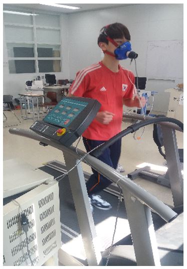
All participants in this study performed sufficient warm-up followed by measurement of maximum oxygen uptake, as shown in (Figure 1). The maximum oxygen uptake was measured after each participant completed treadmill running at a slope of 0° and starting speed of 5.4 km/hr, which was increased by 1.2 km/hr until exhaustion (Korea Institute of Sport Science, 2014). For each participant, the running speed at 70% of the measured maximum oxygen uptake (average, 2.52 ± 0.26 m/sec) was calculated and used for the 65-min running session on a treadmill with two ground reaction force generators (instrumented dual-belt treadmill; Bertec, USA) for inducing fatigue (Wilmore & Costill, 1994).
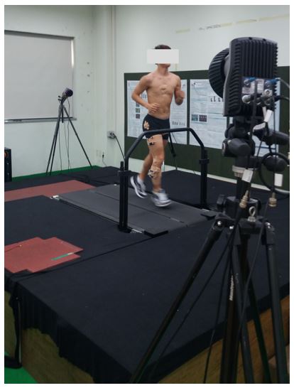
Before the actual run data collection, eight infrared cameras operating at 100 Hz/sec (Oqus 300; Qualisys, Sweden) were installed in the right space of the running participant in order to obtain three-dimensional kinematic data (Figure 2). Camera calibration was performed by using nonlinear transformation (NLT) after fixing an L-shaped frame with four markers of known lengths on the right back side of the treadmill for the global coordinate setting. The axis directions were determined as follows: the upward vertical axis of the global coordinate as +Z, the running direction as +Y, and the right-hand coordinate configuration that takes the cross of +Y onto +Z as +X. The local coordinate to the shank and foot of the right segment had the same directions as the global coordinates. To trace the three-dimensional motion of the two lower-body segments, three non-collinear reflector markers were attached to the side of the shank as well as to the lateral malleolus, the caps of the running shoes, and the fifth metatarsal head skin on the foot segment. One person was in charge of marker attachment in order to minimize the differences in marker applications. Before the run started, kinematic data were collected by using the installed camera for about 3 sec while the participant stood still for the gathering of anatomical data for the calculation of the three-dimensional angle of the lower-body segment. The sampling frequency of the kinematic data collected by the treadmill with the installed ground force reactors was set to 1,000 Hz, and gathered while being synchronized along with the kine- matics data.
Data were collected for at least 20 strides at the 5-min time point after the run started when the body had completely adjusted to running in the non-fatigued[ state, and at the 65-min time point when the body was assumed to be fatigued. The subjects were not aware of the col- lection of data in both cases (Ryu, 2013, 2014). In this study, running shoes with the same cushioning index were provided to all participants.
3. Data processing and analysis
The three-dimensional angle of shank-shank and foot segment motion was collected by using the NLT method and was calculated by using the Cardan method (Hamill & Ryu, 2003). The foot inversion-eversion angle and the shank internal-external rotation angle were selected from the calculated segment movement for the calculation of the coupling angle.
The vector coding technique was used for the coupling angle com- putation (Hamill, Haddad, & McDermott, 2000; Sparrow, Donovan, van Emmerik, & Barry, 1987; Tepavac & Field-Fote, 2001). The internal-external rotation angle of the shank as the proximal segment was set on the x-axis and the inversion-eversion angle of the foot as distal segment on the y-axis, and the direction of this relative movement was determined with the average sine and cosine calculation of each direction (Ferber, Davis, & Williams, 2005; Heiderscheit et al., 2002; Ryu, 2006). Therefore, the absolute addition vector between the two adjacent data points within the stride interval during running was calculated by using the following equation:
Here, i = 1, 2, ..., n.
The analysis interval was set from when the vertical ground reaction force is 5 N or higher during the first stride when the right foot lands on the treadmill to when the same force is less than 5 N during the 20th stride when the right foot leaves the treadmill.
No filtering was done to all kinematic and kinetic data used in this study, as it was assumed that the data used for calculating coupling and free torque can distort the results of nonlinear analysis (Buzzi, Stergiou, Kurz, Hageman, & Heidel, 2003; Kantz & Schreiber, 1997; Ryu, 2006). Free torque was computed to observe the coupling angle due to running fatigue and its kinematic relations. Free torque was computed for the analysis interval as described above, and the magnitude of the free torque was processed as non-dimensional after being standardized by using height and weight. The study by Ryu (2016) was used for a more specific calculation process. The nonlinear approximate entropy (ApEn) technique was used to observe the time series of how running-induced fatigue affects the periodicity change in coupling of the foot inversion-eversion angle and the shank internal-external rotation angle as well as the free torque. ApEn is the logarithmic probability that shows how the definite distance within a set of data points in state space is relatively similar to the next increment. The ApEn value is low for points that are possibly separated by the same distance from the comparison point, whereas it is high for large distances between the data points. In other words, ApEn evaluates repetitiveness (periodicity) by comparing similar patches in m points to similar points in m + 1 points (m / m + 1), re- presented as integers ranging from 0; the higher the value is, the low the periodicity is (Preatoni et al., 2014).
ApEn(m, r, N) uses N input data points u(1), u(2), …, u(N) and the two input parameters m and r. The input parameter m stands for the pattern length for comparison and r is the tolerance error. The following process was performed for the ApEn calculation (Stergiou, 2004).
A vector continuum x(1) was created from u(i) defined by x(i) = [u(i), …, u(i + m - 1)] by using x(N - m - 1), and the distance d[x(i), x(j)] was defined as the largest distance between the vector x(i) and x(j) among all scalar elements. Then, the vector continuum x(1) was used from x(N - m - 1) to create i # N - m + 1.
The values measure the periodicity of the pattern that is similar to the given pattern of window length m within the tolerance error r. This study used r = 0.2 and m = 2 (Stergiou, 2004).
Finally, Φ^m (r) was defined as the average value of 1n in the form of natural logarithm. ApEn is defined as follows.
MatLab (Mathworks Inc., Natick, MA, USA) was used for data pro- cessing in this study.
The processing of ApEn values of segment coupling and free torque calculated by using nonlinear tools did not use descriptive and in- ductive statistics and only used each individual’s exponent magnitude for analysis in order to exclude any possibility that within-subject perio- dicity may be distorted as the unique characteristics of individuals may affect the average (Ryu, 2009). Therefore, the effect of running-induced fatigue on the periodicity of segment coupling and the relationship of the periodicity of the coupling to that of the free torque only considered the magnitude of the ApEn value for the comparison of data before and after fatigue based on the participant rate.
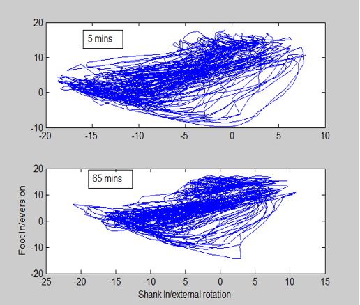
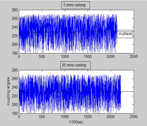
The ApEn analysis results based on the above-described method, to observe how the periodicity changes in the coupling of foot inversion-eversion and shank internal-external rotation and its relationship to the free torque periodicity, are shown in (Table 2). In addition, (Figure 3) shows the shank internal-external rotation angle before and after the induction of fatigue, which was calculated before the calculation of coupling in the foot-shank segment, and (Figure 4) the foot inversion-eversion angle. The angle-angle diagram of these two segment angles necessary for coupling calculation is illustrated in (Figure 5), and the coupling angle of the two segments in (Figure 6).
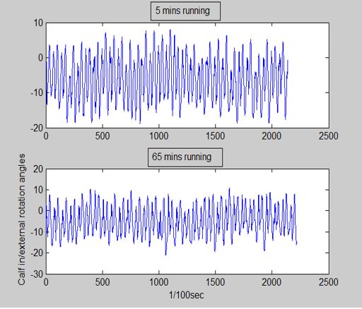
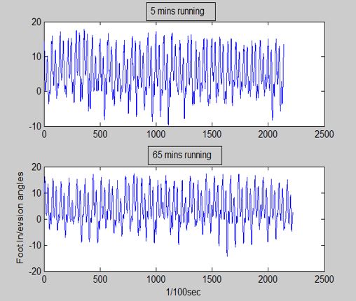
The ApEn value for the coupling of foot inversion-eversion and shank internal-external rotation immediately after 65 min of running (fatigued condition) increased in 12 of 15 participants (80%), which indicates low periodicity. Concerning the periodicities of fatigued and non-fatigued shank internal-external rotation angles and foot inversion-eversion angle, which make up this coupling, the ApEn value of the shank internal- external rotation angles increased in 12 of 15 participants (80%), indi- cating low periodicity, whereas the ApEn value of the foot inversion-eversion angle decreased in 10 of 15 participants (66%), showing high periodicity. Similar to the coupling and the shank internal-external rota- tion angle, free torque also demonstrated reduced periodicity, as the ApEn value increased in 12 of 15 participants (80%). The fluctuation trend of the ApEn value of each participant's coupling and shank internal-external rotation angle in the fatigued and non-fatigued conditions was identical for 10 participants; that is, 10 of 15 participants (66%) exhibited reduction in the coupling periodicity between shank internal-external rotation and foot inversion-eversion, as well as in the periodicity of the shank internal-external rotation. In contrast, like the coupling angle, the periodicity of the foot inversion-eversion to the coupling was low in 5 of 15 participants (33%), after running for 65 min. Thus, the periodicity between the foot inversion-eversion angle and coupling in the fatigued and non-fatigued conditions tended to be less closely related to each other that that between the shank internal-external rotation and coupling. In the examination of each participant to determine how free torque, known to largely affect the foot inversion-eversion movement and the shank internal-external rotation size, is related to their periodicities, 10 of 15 participants (66%) demonstrated reduced periodicity in free torque due to running-induced fatigue or in coupling. Nine of 15 participants (60%) displayed low periodicity, showing a similar tendency of perio- dicity changes in free torque and in shank internal-external rotation. In contrast, only 4 of 15 participants (26%) showed periodicity changes in free torque and in foot inversion-eversion, which are relatively lower than in the other condition.
|
Participant |
Coupling |
Calf internal/external
rotation |
Foot inversion/eversion |
Free torque |
||||
|
5 min |
65 min |
5 min |
65 min |
5 min |
65 min |
5 min |
65 min |
|
|
1 |
1.988 |
2.012 |
0.745 |
0.775 |
0.709 |
0.665 |
1.194 |
1.351 |
|
2 |
1.999 |
1.967 |
0.626 |
0.657 |
0.820 |
0.767 |
1.148 |
1.039 |
|
3 |
1.947 |
1.962 |
0.900 |
0.961 |
0.591 |
0.614 |
1.037 |
0.964 |
|
4 |
2.030 |
2.043 |
0.914 |
0.927 |
1.014 |
0.946 |
1.363 |
1.176 |
|
5 |
1.997 |
2.009 |
0.879 |
0.964 |
0.664 |
0.707 |
1.123 |
1.216 |
|
6 |
1.982 |
1.972 |
0.912 |
0.950 |
0.737 |
0.671 |
1.040 |
1.139 |
|
7 |
1.996 |
2.008 |
0.724 |
0.861 |
0.649 |
0.712 |
1.071 |
1.134 |
|
8 |
1.990 |
2.017 |
0.717 |
0.818 |
0.552 |
0.613 |
1.233 |
1.419 |
|
9 |
1.906 |
1.966 |
0.752 |
0.774 |
0.697 |
0.686 |
1.064 |
1.268 |
|
10 |
1.920 |
1.954 |
0.720 |
0.758 |
0.485 |
0.476 |
1.098 |
1.222 |
|
11 |
1.941 |
2.003 |
0.949 |
0.984 |
0.698 |
0.717 |
1.147 |
1.288 |
|
12 |
1.954 |
1.955 |
1.003 |
0.913 |
0.610 |
0.604 |
1.057 |
1.079 |
|
13 |
1.947 |
1.931 |
0.823 |
0.807 |
0.798 |
0.702 |
1.085 |
1.105 |
|
14 |
1.990 |
2.014 |
1.041 |
0.950 |
0.889 |
0.866 |
0.981 |
1.024 |
|
15 |
1.911 |
1.970 |
0.953 |
0.996 |
0.711 |
0.657 |
1.006 |
1.081 |
Analysis of bodily stability change during human body movements is important in predicting possible injury and fall risks, and in estab- lishing strategic training programs (Ryu, 2014). During motions such as running, stability reflects the sensitivity of the dynamic system to any bodily perturbation. Dynamic stability assessment during running can be done by observing changes in stride-to-stride cycles (Stergiou, 2004). Both linear and nonlinear tools can be used to observe these changes. Linear tools can be limited in clarifying the real structure of movement changes as the processes typically make the decision by applying statis- tical techniques after analyzing a few strides, and if the time function is standardized, any temporary changes in movement pattern is likely to be lost. On the contrary, nonlinear tools focus more on how the motion pattern changes over time (Dingwell & Cusumano, 2000; Hausdorff, Edelberg, & Michell, 1997). Therefore, when observing the stability of a specific variable in running, nonlinear tools can study the changes of stride-to-stride cycles during a longer period, making it advantageous to observing the original conserved data and to analyzing the un- iqueness of a system (Abraham et al., 1986).
In this study, stability was analyzed in the fatigued state during the course of running by using a nonlinear tool, ApEn, on the coupling of foot inversion-eversion and shank internal-external rotation (which is related to injury during exercise) (Hamill & Haddad, 2002), the foot inversion-eversion and the shank internal-external rotation angle, and free torque (which is closely related to the two angles) (Milner et al., 2006; Pohl et al., 2009). The results showed increased ApEn values in shank internal-external rotation, coupling of shank internal-external rota- tion and foot inversion-eversion, and free torque, in 80% of the study participants in the fatigued state after running for 65 min. Such result suggests that fatigue decreased the periodicity of the foot inversion-eversion and the shank internal-external rotation coupling (Nigg et al., 1993, 1998; Ryu, 2004) and that of the free torque (Pohl et al., 2009), inducing an unstable state (Preatoni et al., 2014).
The relative coupling movement of the foot inversion-eversion and the shank internal-external rotation, the overuse of which is known to be related to injury during moving exercises such as running, is one of the traditionally studied topics in sports mechanics related to injury (Ryu, 2004). The movement on foot inversion-eversion occurring in the subtalar joint and the ankle joint (Hintermann, Nigg, Sommer, & Cole, 1994) transfers to shank internal-external rotation, making it closely connected to knee pain (Bahlnen, 1988; James et al., 1978; Ryu, 2004). Nigg et al. (1993) claimed that a runner with knee pain and weak joint force may have poor coordination of shank rotation and rear-foot ever- sion (Ryu, 2004). On the basis of these previous studies, the unstable coupling in the fatigued condition must have largely affected the knee; however, this seems to result from foot-shank coupling attempting to flexibly adapt to the fatigued state (Lipsitz, 2002; Ryu, 2014). One of the previous studies that used a linear tool for periodicity analysis showed that the periodicity of this coupling increased at moments of heel strike and toe off (Ryu, 2004), but this should not be compared with the result of this study owing to the differences in analysis tools and analysis timing (Ryu, 2014).
The study results showed an unstable state of shank internal-external rotation with lowered periodicity in 80% of the participants in the fati gued condition; however, the ApEn value of foot inversion-eversion motion was reduced in 10 of 15 participants (60%), displaying increased periodicity with maintained stability. This seems to be the result of the instinctive strategy to maintain a consistent range of bodily motion because of relatively larger muscular fatigue in foot inversion-eversion (Helbostad, Leirfall, Moe-Nilssen, & Sletvold, 2007). This result also matches with that of a previous study showing a consistently maintained range of foot inversion-eversion in the fatigued condition due to pro- longed running (Ryu, 2001). Such phenomenon, as described above, seems to be the effect of neuromuscular action to adapt to and maintain stability in the fatigued condition (Preatoni et al., 2014); however, it also has the possibility of injury owing to low adaptability to the fatigued condition and low flexibility to such an adaptation (Lipsitz, 2002; Preatoni et al., 2014; Ryu, 2014). This is consistent with previous studies claiming that neurological patients have a higher chance of injury despite their slower walking pace and high local periodicity (Dingwell & Cusumano, 2000; Ryu, 2014). Therefore, to improve the adaptability of foot inversion-eversion to fatigue during prolonged running, the muscular force and muscular endurance of the foot segment must be increased.
Free torque, closely related to shank stress fracture during running (Milner et al., 2006; Pohl et al., 2009), displayed reduced periodicity in many participants in the fatigued condition, as the ApEn value appeared large as in coupling. According to the study by Ryu (2016), the instant- aneous absolute free torque at the maximum braking force at the landing moment is larger after running for 5 min than for 125 min, showing a decreasing trend of free torque in the fatigued state. This shows that prolonged running-induced fatigue rather reduces the absolute magnitude of free torque at the moment of braking and does not largely cause shank stress fracture. The decreased periodicity of free torque in this study also seems to indicate the same tendency as its absolute magnitude concerning the potential risk.
It has been reported that fatigue induced by prolonged running affects each movement involving the lower body (Brüggemann et al., 1994; Ryu, 2001). Injuries from prolonged running would result more from chronic causes due to the accumulation of impulse force than from acute causes due to a large impulse force in one moment. A time-series analysis of how bodily movement changes owing to excessive use of the human body is necessary to diagnose the cause of injury from prolonged running. In particular, it is important to analyze the periodicity of the relative angle of the mutual coupling movement in the shank segment, which is known to be connected to injury during running, to predict the potential risk of lower-body injury. Moreover, the periodicity change in free torque over time in the fatigued state induced by prolonged running also needs to be studied to investigate its relation to stability. In this study, 66% of the participants exhibited reduced periodicity of coupling and free torque due to the prolonged running-induced fatigue, and 60% showed decreased periodicity in shank internal-external rotation and free torque. However, a very low percentage of participants showed the same trend in changes of foot inversion-eversion periodicity and free torque periodicity. Such results, as explained above, suggested that many subjects manifested consistency in foot inversion-eversion periodicity unlike in the other variables. This study analyzed the periodicities of the coupling movement between the foot inversion-eversion and the shank internal-external rotation and free torque, and demonstrated reduced periodicity due to running fatigue in coupling, shank internal-external rotation angle, and free torque, but increased periodicity in foot inversion-eversion. Therefore, the potential risk of injury is most likely attributable to foot inversion-eversion while running (Preatoni et al., 2014).
This study analyzed the periodicity of the coupling angle between shank internal-external rotation and foot inversion-eversion and that of free torque, which is known to be closely related to the movement of these angles, in 15 male participants in their late 20s with a rear-foot strike, after running for 5 and 65 min at 70% of their maximum oxygen uptake. The analysis results showed reduced periodicity in 80% of the participants due to fatigue in shank internal-external rotation, coupling of shank internal-external rotation and foot inversion-eversion, and free torque. The change in the periodicity of free torque before and after the running-induced fatigue and the periodicity of foot inversion-eversion showed a similar trend in 66% of the participants, and the change in the periodicity of free torque and the shank internal-external rotation angle in 60%. On the basis of these results, the following con- clusions can be made. The foot inversion-eversion movement in the fatigued state due to prolonged running has a relatively lower flexibility to adapt to the fatigued condition than the coupling between the foot inversion-eversion and the shank internal-external rotation, the move- ment of shank internal-external rotation, and free torque; thus, it may be conjectured that the risk of potential injury is higher. For follow-up studies, more subjects may be needed to overcome the limitations in generalization, and techniques such as a surrogate data test can be explored for better reliability of the analysis.
References
1. Abraham, N. B., Albano, A. M., Das, B., Guzman, G. D., Yong, S., Gipggia, R. S., Puccioni, G. P. & Tredicce, J. R. (1986). Calculating the dimension of attractors from small data sets. Physics Letters, 114A(5), 217 -221.
Crossref
Google Scholar
2. Bahlnen, A. (1988). The etiology of running injuries: a longitudinal, prospective study. Ph.D. thesis, The university of Calgary, calgary, Alberta.
Crossref
3. Boyer, K. A., Silvernail, J. F. & Hamill, J. (2014). The role of running mileage on coordination patterns in running. Journal of Applied Biomechanics, 30, 649-654.
Crossref
Google Scholar
PubMed
4. Buzzi, U. H., Stergiou, N., Kurz, M., Hageman, P. A. & Heidel, J. (2003). Nonlinear dynamics indicates aging affects variability during gait. Clinical Biomechanics, 18(5), 435-443.
Crossref
Google Scholar
5. Brüggemann, G. P., Arndt, A., Kersting, U. G. & Knicker, A. J. (1995). Influence of fatigue on impact force and rearfoot motion during running. Proceedings of International Society of Biomechanics, 132 -133.
Crossref
6. Choi, E. S. & Song, M. S. (2003). Concept analysis: fatigue. Korean Journal of Woman Health Nursing, 9(1), 61-69.
Crossref
Google Scholar
7. Clement, D. B., Taunton, J. E., Smart, G. W. & McNicol, K. L. (1981). A survey of overuse running injuries. Medicine & Science in Sports & Exercise, 13(2), 83.
Crossref
8. Collins, J. & Whittle, M. W. (1989). Impulsive forces during walking and their clinical implications. Clinical Biomechanics, 4(3), 179-187.
Crossref
Google Scholar
PubMed
9. Dingwell, J. B. & Cusumano, J. P. (2000). Nonlinear time series analysis of normal and pathological human walking. Chaos, 10(4), 848-863.
Crossref
Google Scholar
PubMed
10. Elliot, B. & Ackland, T. (1981). Biomechanical effects of fatigue on 10,000 meter running technique. Research Quarterly for Exercise and Sport, 52(2), 160-166.
Crossref
Google Scholar
PubMed
11. Ferber, R., Davis, I. M. & Williams III, D. S. (2005). Effect of foot orthotics on rearfoot and tibia joint coupling patterns and variability. Journal of Biomechanics, 38(3), 477-483.
Crossref
Google Scholar
PubMed
12. Gibson, H. & Edwards, R. H. T. (1985). Muscular exercise and fatigue. Sports Medicine, 2(2), 120-132.
Crossref
Google Scholar
PubMed
13. Hamill, J. & Haddad, J. M. (2002). The role of variability in the etiology of running. Proceedings of 2002 KNUPE International Symposium, 2(1), 107-18.
Crossref
14. Hamill, J., Haddad, J. M. & McDermott, W. J. (2000). Issues in quantifying variability from a dynamical systems perspective. Journal of Applied Biomechanics, 16(4), 407-418.
Crossref
Google Scholar
15. Hamill, J. & Ryu, J. S. (2003). Experiment in sport biomechanics. Daehanmedia, 99-104.
Crossref
16. Hausdorff, J. M., Edelberg, H. K. & Mitchell, S. L. (1997). Increased gait unsteadiness in community dwelling elderly fallers. Archives of Physical Medicine and Rehabilitation, 78(3), 278-283.
Crossref
Google Scholar
PubMed
17. Heiderscheit, B. C., Hamill, J. & Van Emmerik, R. E. A. (2002). Variability of stride characteristics and joint coordination among individuals with unilateral patellofemoral pain. Journal of Applied Biomechanics, 18(2), 110-121.
Crossref
Google Scholar
18. Helbostad, J. L., Leirfall, S., Moe-Nilssen, R. & Sletvold, O. (2007). Physical fatigue affects gait characteristics in older persons. The Journals of Gerontology Series A : Biological Sciences and Medical Sciences, 62(9), 1010-1015.
Crossref
Google Scholar
19. Hintermann, B., Nigg, B. M., Sommer, C. & Cole, G. K. (1994). Transfer of movement between calcaneus and tibia in vitro. Clinical Biomechanics, 9(6), 349-355.
Crossref
Google Scholar
PubMed
20. Holden, J. P. & Cavanagh, P. R. (1991). The free moment of ground reaction in distance running and its changes with pronation. Journal of Biomechanics, 24(10), 887-897.
Crossref
Google Scholar
PubMed
21. James, S., Bates, B. & Osternig, L. (1978). Injuries in running. The American Journal of Sports Medicine, 6(1), 40-50.
Crossref
PubMed
22. James, R., Dufek, J. S. & Bates, B. T. (1992). Effects of fatigue on mechanical and muscular components of performance during drop landings. Proceedings of NACOBII :553-554.
Crossref
23. Jean-Benoit, M., Pierre, S. & Guillaume, Y. H. (2011). Changes in running kinematics, kinetics, and spring-mass behavior over a 24-h run. Medicine & Science in Sports & Exercise, 43(5), 829-836.
Crossref
Google Scholar
PubMed
24. Kantz, H. & Schreiber, S. (1997). Nonlinear time series analysis. Cambrige University Press, Cambridge, UK.
Crossref
Google Scholar
25. Korea Institute of Sport Science [KISS] (2014). Report for Model Development of Fitness Managing Center.
Crossref
26. Lipsitz, L. A. (2002). Dynamics of stability: The physiologic basis of functional health and frailty. Journal of Gerontology : Biological Sciences, 57A(3), B115-B125.
Crossref
Google Scholar
PubMed
27. McFarland, P. A. (1971). Understanding fatigue in modern life. Ergonomics, 14(1), 1-10.
Crossref
Google Scholar
PubMed
28. Milner, C. E., Davis, I. S. & Hamill, J. (2006). Free moment as predictor of tibial stress fracture in distance runners. Journal of Biomechnics, 39(15), 2819-2825.
Crossref
Google Scholar
PubMed
29. Nigg, B. M., Cole, G. & Nachbauer, W. (1993). Effects of arch height of the foot on angular motion of the lower extremities during running. Journal of Biomechanics, 26(8), 909-916.
Crossref
Google Scholar
PubMed
30. Nigg, B. M., Khan, A., Fisher, V. & stefanyshyn, D. (1998). Effect of shoe insert construction on foot and leg movement. Medicine Science Sports Exercise, 30(4), 550-555.
Crossref
Google Scholar
PubMed
31. Pohl, M. B., Hamill, J. & Davis, I. S. (2009). Biomechanical and anatomical factors associated with a history of plantar fasciitis in female runners. Clinical Journal of Sports Medicine, 19(5), 372-376.
Crossref
Google Scholar
32. Preatoni, E., Hamill, J., Harrison, A. J., Hayes, K., Emmerik, V. R., Wilson,C. & Rodano, R. (2014). Movement variability and skills monitoring in sports. Sports Biomechanics, 12(2), 69-92.
Crossref
Google Scholar
PubMed
33. Ryu, J. S. (2001). Fatigue effects on biomechanical parameters during a prolonged run. The Korean Journal of Physical Education, 40(4), 1011-1025.
Crossref
34. Ryu, J. S. (2004). The effect of fatigue caused by a prolonged run on the variability of the lower extremity segment coupling. The Korean Journal of Physical Education, 43(3), 803-811.
Crossref
35. Ryu, J. S. (2006). The elderly's coupling pattern between the foot and the tibia and its variability during walking. The Korean Journal of Physical Education, 45(1), 747-756.
Crossref
36. Ryu, J. S. (2009). The effect of walking with high-heel shoes on local dynamic stability.The Korean Journal of Physical Education, 48(1), 431-438.
Crossref
37. Ryu, J. S. (2013). Effect of a prolonged-run-induced fatigue on the ground reaction force components. Korean Journal of Sport Biomechanics, 23(3), 225-233.
Crossref
Google Scholar
38. Ryu, J. S. (2014). Variability of GRF components between increased running times during prolonged run. Korean Journal of Sport Biomechanics, 24(4), 359-365.
Crossref
Google Scholar
39. Ryu, J. S. (2016.) Effect of prolonged running-induced fatigue on freetorque components. Korean Journal of Sport Biomechanics, 26(1), 31-37.
Crossref
Google Scholar
40. Sparrow, W. A., Donovan, E., van Emmerik, R. E. A. & Barry, E. B. (1987). Using relative motion plots to measure changes in intra-limb and inter-limb coordination. Journal of Motor Behavior, 19(1), 115-129.
Crossref
Google Scholar
41. Stergiou, N. (2004). Innovative analyses of Human Movement, Human Kinetics, 76-84.
Crossref
42. Tepavac, D. & Field-Fote, E. C. (2001). Vector coding: A technique for quantification of intersegmental coupling in multicyclic behaviors. Journal of Applied Biomechanics, 17(3), 259-270.
Crossref
43. Wilmore, J. H. & Costill, D. L. (1994). Physiology of sport and exercise. Human kinetics, 110-522.
Crossref
Google Scholar
