Open Access, Peer-reviewed
eISSN 2093-9752
Open Access, Peer-reviewed
eISSN 2093-9752
Ja Yeon Lee
Chae Kwan Lee
Shuho Kang
Il Bong Park
https://dx.doi.org/10.5103/KJAB.2023.33.4.119 Epub 2023 November 19
Abstract
Objective: This study aimed to compare the activity of the trunk and leg muscles while performing fente (in fencing) wearing weighted and waterbag vests.
Method: The electromyography test was used to measure and analyze the activation of the trunk and leg muscles. Eight active fencers from B University (age: 19.5 ± 0.66 years, height: 179.75 ± 5.93 cm, weight: 72 ± 6.32 kg) were selected for this study.
Results: According to the EMG analysis results of the 4 muscles measured in this study, left-right differences were observed for rectus abdominis and external oblique abdominis, but left-right differences between the groups were not significant. The gluteus medius muscle was not significantly different from the adductor muscle, but there were significant differences between the groups.
Conclusion: The electromyographic analysis of the four muscles measured in this study revealed no significant difference between the left and right recti abdominis and external obliques depending on the vests. However, significant differences were observed between the left and right gluteus medius and adductor longus. Our results can be interpreted as the effects of the inherent movements involved in the fente. Furthermore, our results indicate that the weight transfer while wearing a waterbag vest, which provides an unstable environment, increased the activity of leg muscles.
Keywords
Electromyography Instability Waterbag vest Fencing athletes
Fencing matches are played on pistes of 1.8 and 2 m wide and 14 m long. Strength, power, agility, speed, and reaction time are the fitness factors associated with fencing performance, as the first fencer to touch the other scores (Barth & Beck, 2007; Roi & Bianchedi, 2008). Due to its unique asymmetrical movements, fencing places high physiological demands on neuromuscular control, strength, and the musculoskeletal system (Murgu, 2006). In addition, the rapid offensive and defensive movements to score points against the opponent expose fencers to varying degrees of impact and force distributed asymmetrically throughout their bodies (Sinclair, Bottoms, Taylor & Greenhalgh, 2010). Accordingly, it is believed that training in sport-specific methods is more necessary given the asym- metrical characteristics of fencing.
Resistance training improves the ability to generate muscle strength under specific conditions (Aspenes & Karlsen, 2012) and increases the speed at which strength is enhanced by increasing maximum strength (Suchomel, Nimphius & Stone, 2016). Athletes gained over 10% of their athletic ability in a study of an unconventional training method of wearing a weighted vest during training (Khlifa et al., 2010). Additionally, Khlifa et al. (2010) reported that adult male basketball players who performed plyometric exercises while wearing a weighted vest over 10 weeks of training improved vertical jump ability more than those who performed plyometric exercises without wearing a weighted vest.
Instability-based resistance training has been proposed as a method for neuromuscular stress adaptation than trad- itional resistance training (Behm & Anderson, 2006). Performing resistance exercises in unstable conditions promoted the activation of stabilizing muscles, thereby amplifying the effectiveness of training (Nairn, Sutherland & Drake, 2017). Further- more, using instability tools in functional strength training improved neuromuscular adaptation and strength enhance- ment (Saeterbakken & Fimland, 2013; Ditroilo et al., 2018). Additionally, neuromuscular training induced through training using agility and instability has been reported to improve core stability (Araujo, Cohen & Hayes, 2015). Training using in- stability tools (e.g., Swiss ball, BOSU, wobble board, air cushion, and slash pipe) has primarily been performed in therapeutic settings. However, this type of training improves the effects of training and is now primarily used in the training of athletes (Behm & Colado, 2013; Chance-Larsen, Littlewood & Garth, 2010; Fowles, 2010).
Several previous studies have investigated the effectiveness of neuromuscular training for muscle activation using instability tools, and these studies focused on the activation of the trunk muscles by rendering the support surface unstable using instability tools, such as a Swiss ball or BOSU. The activation of the leg and trunk muscles is substantially affected by the use of instability tools to maintain balance and stability during lifting (Kohler, Flanagan & Whiting, 2010; Li, Cao & Chen, 2013; McBride, Larkin, Dayne, Haines & Kirby, 2010). While there are several methods for using instability tools, studies on exercises on an unstable surface that are focused on athletic movement during the sport are insufficient. Training methods that include instability can strengthen the athlete's training system according to the principle of the specificity of the sport, and athletes are encouraged to practice athletic movements of the sport in the same environment where they will compete (Behm & Anderson, 2006).
A water bag has the structure of inflatable tubes resembling barbells with water and is mainly used for functional strength training (Wezenbeek et al., 2022). When using the water bag, the water in the tubes produces small but rapid vibrations, and these vibrations prompt the user to maintain their balance constantly (Nairn, Sutherland & Drake, 2015). Water bag devel- opers and researchers state that training using this tool can activate the user's torso muscles.
Wezenbeek et al. (2022) investigated the effect of using water bags and wooden sticks during three functional exercises, squats, lunges, and step-ups, on the activities of the muscles of the trunk and lower extremities. The abdominal and gluteal muscle activities increased considerably during the three ex- ercises. Additionally, Calatayud et al. (2015) analyzed the activity of the trunk muscles during clean and jerk movements using barbells, water bags, and sandbags, and reported that the activity of the trunk muscles was high when using water bags.
Core muscle stability in a sports environment is the ability to control the position and movement of the trunk to optimally generate, transmit, and control force and movement in an integrated exercise environment (Kiber, Press & Sciascia, 2006).
Muscle activation measured by electromyography (EMG) plays an important role in all motor activities because it is assumed to originate from postural activation mechanisms (Borysiuk, Blaszczyszyn, Piechota, Konieczny & Cynarski, 2022). Witkowski et al. (2018) reported on the importance of core muscles that maintain posture based on the finding that the back and abdominal muscles were activated first in a study on wheelchair fencers. Additionally, Borysiuk et al. (2020) and Kriventsove et al. (2017) reported that postural muscles that stabilize the trunk play a critical role due to the dynamism of attack techniques during fencing matches.
Electromyography (EMG) testing is useful in assessing muscle activation (Ghezelbash, EI Ouaaid, Shirazi-Adl, Plamondon & Arjmand, 2018), and a positive relationship between muscle activation and performance in sports can be established (Lynn, Watkins, Wong, Balfany & Feeney 2018).
Research on resistance training has shown that if the goal is simply to increase core muscle activation, lifting heavy loads on a stable base of support may be sufficient (Anderson, Gaetz, Holzmann & Twist, 2013). However, this study aims to study the sport-specific aspects of fencing based on previous research showing that training through instability can con- tribute to neuromuscular improvement as well as increased strength (Cowley, Swensen & Sforzo, 2007). When comparing dynamic muscle activation during fencing movements using a weight vest and a water bag vest, it is believed that dynamic muscle activation will be higher when using a water bag vest.
In Korea and overseas, research on specific movements in sports with asymmetric movements, such as fencing, is still insufficient. In addition to the paucity of studies on the effects of using water bags, there is a lack of studies on the sport-specific, instability-related characteristics that are unique to each sport.
Therefore, this study aimed to provide basic data that can aid the efficient performance of the fente movement in fencing by using weighted and waterbag vests. Our findings contribute to improving the fencers' performance.
1. Participants
Eight active male athletes (height: 179.75±5.93, weight: 72±6.32) from the B University fencing team in B Metropolitan City participated in this study. Athletes with current musculo- skeletal injuries and those who have not recovered were excluded from the competition.
For the number of subjects in this study, the G Power 3.0 program (Faul, Erdfelder, Lang & Buchner, 2007) was applied with an effect size of 0.5, power of 0.8, and significance level of 0.05. There were 10 subjects who voluntarily agreed to participate in the study, but 8, excluding 2 due to injuries, were selected as final research subjects.
(Table 1) shows the physical characteristics of the partici- pants. The study was conducted after explaining the aim to all participants and obtaining their consent voluntarily.
|
|
Age (yrs.) |
Height (cm) |
Weight (kg) |
Career (yrs.) |
|
Participants (n=8) |
19.5±0.66 |
179.75±5.93 |
72±6.32 |
7.75±1.19 |
|
Values are Mean ± SD |
||||
2. EMG Measurement methods and instruments
1) Surface EMG device and attachment sites
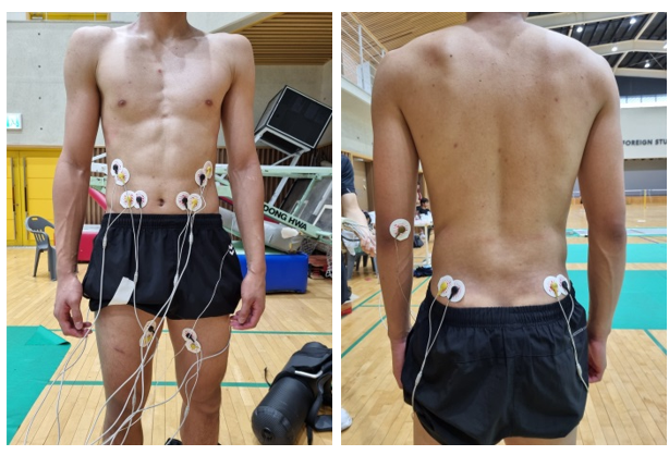
Fencing is a unilateral exercise performed in an asymmetric position while holding a sword in one hand, resulting in strength imbalances in the legs due to the different roles of the front and back legs, and these changes can also be observed in the torso muscles (Guilhem, Giroux, Couturier, Chollet & Rabita, 2014). In this study, the activities of rectus abdominis, external oblique, gluteus medius, and adductor longus were measured to compare the activities of the trunk and leg muscles in relation to the exercise equipment used (Figure 1). The semi-wireless surface EMG device WEMG8 (Lextha, Korea, gain = 1,000, input impedance > 1012, CMRR > 100 dB) was used for the measurement. The sampling frequency was set to 1,024 Hz, and the Telescan (Laxtha, 2019) program was used. Before the electrode attachment, the attachment site was cleaned with an alcohol-based disinfectant using cotton wipes to minimize skin resistance. For the EMG analysis, the maximum voluntary isometric contraction (MVIC) was measured and set to the corresponding % MVIC value.
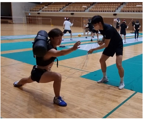
Maximal voluntary isometric contraction is measured for individual muscles when exerting maximum force against isometric resistance using the Manual Muscle Testing (MMT) method of Daniels and Worthingham's muscle testing (Brown, Hislop & Avers, 2013). Data were collected three times for 5 seconds each. Rectus abdominis was measured by applying resistance to the flexor muscles in a supine position, with the lower limbs fixed, and the trunk flexed until the scapula came off the floor. The External Oblique was measured by applying resistance with the trunk rotated in a position with the knees up and the trunk flexed. Gluteus Medius was measured in a lying position with the hips and knees placed to the side and the hips abducted 10°. The adductor longus was measured by applying resistance with the lower leg adducted while lying down with the hips and knees placed to the side. Active and reference electrodes were attached to the most developed parts of the belly of the muscle, 1 cm away in parallel to the direction of the muscle fibers (Badier, Guillot, Lagier-Tessonnier, Burnet & Jammes, 1993). In addition, assistants moved the EMG device together to minimize participant discomfort and prevent motion artifacts during the movements (Figure 2). (Table 2) shows the experimental devices.
|
Equipment |
Model |
Manufacture |
|
EMG equipment |
WEMG8 |
Laxtha.
Korea |
|
EMG analysis |
Telescan |
Laxtha. Korea |
|
Surface |
AG/AgCI
2223 |
3M.
Korea |
|
AG : Silver |
||
2) EMG Data collection interval
This study was conducted to verify the difference in muscle activities between the tools used to train the fente movement of fencers. The fente movement was performed three times on signal while wearing each tool. The movement was per- formed three times while wearing a weighted vest and three times with a waterbag vest, with a 3 min rest period between each set. The participants stopped all movements as soon as the fente foot touched the ground, and data was collected from the moment the foot touched the ground until 1 s after. All movements were synchronized to a 3-s interval, and the participants were instructed to perform the movements in time with the sound signal of the metronome in the computer program. After a sufficient explanation of the sound signal and timing, the participants were given multiple opportunities to adapt to the movement. The means of the measurements collected three times for each instrument were calculated.
3. Data analysis
All EMG data measured in this experiment were processed using a band-pass filter of 20 to 450 Hz, and the values converted into root mean square (RMS) were used. The MVICs of the muscles for measurement were measured before the start of the experiment, and the muscle activation during fente movement was normalized using the previously measured MVIC and the following equation.
where, : RMS value of muscular activation during the movement
: Mean RMS of muscle activation at maximum voluntary isometric contraction
4. Statistical analysis
All collected data are presented as means and standard deviations. These data were calculated using SPSS 28.0. The Shapiro-Wilks test was used to analyze the normality of all general characteristics and variables. A paired t-test was per- formed to verify the difference between the left and right sides of a muscle group, and an independent-sample t-test was performed to confirm the differences between the left and right muscles between the experimental groups. P < .05 was considered to be statistically significant.
(Table 3) shows the differences in EMG results of the fente movement between muscle groups.
|
Variable |
Group |
Right |
Left |
t |
p |
|
Rectus abdominis |
Weight vest |
10.85±5.14 |
7.32±6.21 |
1.487 |
0.181 |
|
Waterbag vest |
17.92±9.20 |
13.65±11.64 |
1.753 |
0.123 |
|
|
t |
-1.896 |
-1.357 |
|
|
|
|
p |
0.079 |
0.203 |
|
|
|
|
External oblique |
Weight vest |
12.15±8.93 |
9.02±2.50 |
1.015 |
0.344 |
|
Waterbag vest |
18.91±18.78 |
13.28±5.70 |
0.869 |
0.414 |
|
|
t |
-0.920 |
-1.939 |
|
|
|
|
p |
0.379 |
0.082 |
|
|
|
|
Gluteus medius |
Weight vest |
12.18±5.40 |
13.11±5.44 |
-1.998 |
0.086 |
|
Waterbag vest |
19.50±4.03 |
20.13±4.28 |
-1.138 |
0.292 |
|
|
t |
-3.074 |
-2.863 |
|
|
|
|
p |
0.009* |
0.013* |
|
|
|
|
Adductor longus |
Weight vest |
16.88±10.94 |
16.59±7.51 |
0.107 |
0.918 |
|
Waterbag vest |
32.80±17.40 |
35.51±11.96 |
-0.609 |
0.562 |
|
|
t |
-2.190 |
-3.789 |
|
|
|
|
p |
0.049* |
0.003* |
|
|
|
|
Values are Mean ± SD, *: p < .05 |
|||||
The activation of rectus abdominis was 10.85±5.14% and 7.32±6.21% for the right and left side, respectively, in the weighted-vest group; however, the difference between the two sides was non-significant. The right and left side of the water- bag vest group was calculated at 17.92±9.20%, and 13.65±11.64%, respectively, and the difference between the left and right was also non-significant. Significant differences were observed between groups regarding the right and left rectus abdominis. In addition, there was a difference between the left and right sides between the weight vest and the water bag vest group, but there was no significant difference.
The activation of external obliques was 12.15±8.93% and 9.02±2.50% for the right and left, respectively, in the weighted-vest group; however, the difference between the two sides was non-significant. For the waterbag-vest group, the muscle activation was 18.91±18.78% and 13.28±5.70% for the right and left side, respectively, with no significant difference be- tween the two. Significant differences were observed between groups for the right and left external obliques. In addition, there was a difference between the left and right sides between the weight vest and the water bag vest group, but there was no significant difference.
The activation of gluteus medius was 12.18±5.40% and 13.11±5.44% for the right and left side, respectively, for the weighted-vest group, with no significant difference between the two sides. In the waterbag vest group, the muscle activation was 19.50±4.03% and 20.13±4.28% for the right and left side, respectively, with no significant difference between the two sides. However, there was a significant difference between the left and right sides of the weight vest and water bag vest groups (p < .05).
The activation of the adductor longus was 16.88±10.94% and 16.59±7.51% for the right and left side, respectively, in the weighted-vest group, with no significant difference between the two sides. In the waterbag-vest group, the muscle activation was 32.80±17.40% and 35.51±11.96% for the right and left side, respectively, with the difference being non-significant. However, there was a significant difference between the left and right sides of the weight vest and water bag vest groups (p < .05).
An important technical aspect of fencing is the fente, a fundamental movement in which the fencer thrusts to gain the upper hand in the attack. The scoring in fencing can be decided by a split of 1/25 s, and in the fente, an attack move- ment requires bold and quick judgment (Roi & Bianchedi, 2008). There are various approaches to training to improve such important fente movements, but the most effective way to improve the movement is by performing them. Herein, muscle activation during a fente movement while wearing weighted and waterbag vests was measured, and the extent to which these tools could activate the fente-specific muscle was examined. While a weighted vest is a tool that increases the body weight and increases movement load, a waterbag vest increases the load for the movement and induces the wearer to maintain balance through the rapid vibration of water in the tubes (Nairn et al., 2015).
Nairn et al. (2015) reported that training with water bags could activate the trunk muscles, and Calatayud et al. (2015) found high trunk muscle activity in the waterbag group while performing clean and jerk.
However, the results of this study were inconsistent with those of previous studies. The activation of the trunk muscles, the rectus abdominis, and external obliques, did not differ significantly between the left and right side depending on the tool used; however, there were significant differences between the left and right leg muscles, the gluteus medius and adductor longus, depending on the tool.
The above results are interpreted as the result of the unique movement characteristics of fencing and the various demands placed on the body during sports activities.
It is necessary to make an approach at high speed and spread the legs wide to thrust the opponent with a saber. The back leg needs to be extended, and the front knee needs to be bent to bring the saber closer to the opponent to execute the above (Thompson, Chang, Alaia, Jazrawi & Gonzalez-Lomas, 2022).
In other words, fencing is a dynamic movement that involves moving the body weight rather than a static movement of the legs in place, and in an actual fencing match, attack and defense techniques must be progressed according to the opponent's movements.
Core muscle activity was numerically different, but unlike the hypothesis, there was no statistically significant difference. McGill (2010) reported that core muscles play a role in maintaining posture during exercise, and lower limb muscles should be mainly used in movements that require speed or power. Based on these previous studies, it is believed that the leg muscles are more involved in the fente movement in fencing because it is dynamic and is a movement for speed and power. Additionally, because leg muscle activity increases in a dynamic posture compared to a static posture in an unstable environment (Park et al., 2015). It is judged that when using a water bag vest that provides an unstable environment, leg muscle activity increased due to strong weight transfer.
The gluteus medius is a typical hip abductor muscle that is concentrically activated when legs are spread apart and eccentrically activated when a single leg is supported (Floyd & Thompson, 2009). The adductor longus provides an intense extensor moment during hip extension and is also active during hip flexion (Neumann, 2010). In this study, when executing the fente movement, the left and right sides of these two muscles were activated simultaneously.
To score against an opponent, fencers must rapidly thrust their weapon at the opponent, which requires explosive force in the back leg to perform a strong forward lunge (Sinclair et al., 2010). Therefore, it is believed the left and right gluteus medius and adductor longus are activated because the fente is an attack motion generated by extending the hind leg at a fast speed while supporting the force with the front leg. In addition, since the fente has the advantage of enabling a faster return to a stable position for defense than other attack methods (Borysiuk & Waskiewicz, 2008), both the left and right muscles are believed to be simultaneously activated as a strategy to prepare for the defense at the same time as the attack.
Fencers have high leg injury rates because fencing requires dynamic and repetitive movements, and the most common injury site is reported to be the knees (19.6%), followed by the thighs (15.2%) and ankles (13.0%) (Wild et al., 2001).
Fencing matches require the ability to respond quickly and appropriately to an opponent's actions and the ability to repeatedly attack and defend (Turner et al., 2014). In particular, the most frequently used attack movements in fencing, such as lunges, are achieved through coordination of the kinetic chain of the ankle, knee, and hip joints (Bottoms, Greenhalgh & Sinclair, 2013). Therefore, it is known that activating lower extremity muscles is helpful for quick transition from offensive movements such as lunges to defensive movements and for preventing leg injuries (Turner et al., 2014).
Accordingly, wearing a waterbag vest when performing fente movements will help improve leg strength and prevent leg injuries.
The purpose of this study was to compare and analyze the activity of the torso and leg muscles according to the weight vest and the water bag vest during fencing fant movements to find out how the torso and leg muscles affect stability during specific movements depending on the tool. The same conclusion was obtained. The results were as follows. Among the four muscles observed, no difference was observed be- tween the left and right sides of the gluteus medius and adductor longus when using a single tool; however, there were significant differences between the left and right sides of the muscles between the tools. Different results from previous studies were obtained due to the unique movement char- acteristics of fencing. However, as the leg muscle activity was high when wearing a water bag vest, It was also found to contribute to improvement training through instability such as a water bag vest not only increases muscle strength but also increases nerve muscle activity.
References
1. Anderson, G. S., Gaetz, M., Holzmann, M. & Twist, P. (2013). Comparison of EMG activity during stable and unstable push-up protocols. European Journal of Sport Science, 13(1), 42-48. https://doi.org/10.1080/17461391.2011.577240
Google Scholar
2. Araujo, S., Cohen, D. & Hayes, L. (2015). Six weeks of core stability training improves landing kinetics among female capoeira athletes: A pilot study. Journal of Human Ki-netics, 45, 27. https://doi.org/10.1515/hukin-2015-0004
Google Scholar
3. Badier, M., Guillot, C., Lagier-Tessonnier, F., Burnet, H. & Jammes, Y. (1993). EMG power spectrum of respiratory and skeletal muscles during static contraction in healthy man. Muscle & Nerve: Official Journal of the American Association of Electrodiagnostic Medicine, 16(6), 601-609. https://doi.org/10.1002/mus.880160605
Google Scholar
4. Barth, B. & Beck, E. (Eds.). (2007). The complete guide to fencing. Meyer & Meyer Verlag.
Google Scholar
5. Behm, D. G. & Anderson, K. G. (2006). The role of instability with resistance training. The Journal of Strength & Con- ditioning Research, 20(3), 716-722. https://doi.org/10.1519 /r-18475.1
Google Scholar
6. Behm, D. G. & Colado Sanchez, J. C. (2013). Instability resistance training across the exercise continuum. Sports Health, 5(6), 500-503. https://doi.org/10.1177/1941738113477815
Google Scholar
7. Borysiuk, Z., Nowicki, T., Piechota, K., Błaszczyszyn, M., Konieczny, M. & Witkowski, M. (2020). Movement patterns and sensori-motor responses: comparison of men and women in wheelchair fencing based on the Polish Paralympic team. Archives of Budo, 16, 19-26.
Google Scholar
8. Borysiuk, Z., Blaszczyszyn, M., Piechota, K., Konieczny, M. & Cynarski, W. J. (2022). Correlations between the EMG Structure of Movement Patterns and Activity of Postural Muscles in Able-Bodied and Wheelchair Fencers. Sensors, 23(1), 135. https://doi.org/10.3390/s23010135
Google Scholar
9. Bottoms, L., Greenhalgh, A. & Sinclair, J. (2013). Kinematic determinants of weapon velocity during the fencing lunge in experienced épée fencers. Acta of Bioengineering and Biomechanics, 15(4), 109-113. https://doi.org/10.5277/ abb130414
Google Scholar
10. Brown, M., Hislop, H. & Avers, D. (2013). Daniels and Worthing- ham's muscle Testing-E-Book: Techniques of manual exa- mination and performance testing. Elsevier Health Sciences.
Google Scholar
11. Calatayud, J., Colado, J. C., Martin, F., Casaña, J., Jakobsen, M. D. & Andersen, L. L. (2015). Core muscle activity during the clean and jerk lift with barbell versus sandbags and water bags. International Journal of Sports Physical Therapy, 10(6), 803. PMID:26618060 PMCID:PMC4637915
Google Scholar
12. Chance-Larsen, K., Littlewood, C. & Garth, A. (2010). Prone hip extension with lower abdominal hollowing improves the relative timing of gluteus maximus activation in relation to biceps femoris. Manual Therapy, 15(1), 61-65. https://doi.org/10.1016/j.math.2009.07.001
Google Scholar
13. Cowley, P. M., Swensen, T. & Sforzo, G. A. (2007). Efficacy of instability resistance training. International Journal of Sports Medicine, 829-835. https://doi.org/10.1055/s-2007-964893
Google Scholar
14. Ditroilo, M., O'Sullivan, R., Harnan, B., Crossey, A., Gillmor, B., Dardis, W. & Grainger, A. (2018). Water-filled training tubes increase core muscle activation and somatosensory con- trol of balance during squat. Journal of Sports Sciences, 36(17), 2002-2008. https://doi.org/10.1080/02640414.2018. 1431868
Google Scholar
15. Faul, F., Erdfelder, E., Lang, A. G. & Buchner, A. (2007). G* Power 3: A flexible statistical power analysis program for the social, behavioral, and biomedical sciences. Behavior Research Methods, 39(2), 175-191. https://doi.org/10.3758 /bf03193146
Google Scholar
16. Floyd, R. T. & Thompson, C. W. (2009). Manual of structural kinesiology (Vol. 16). New York, NY: McGraw-Hill.
Google Scholar
17. Fowles, J. R. (2010). What I always wanted to know about in- stability training. Applied Physiology, Nutrition, and Meta- bolism, 35(1), 89-90. https://doi.org/10.1139/h09-134
Google Scholar
18. Ghezelbash, F., El Ouaaid, Z., Shirazi-Adl, A., Plamondon, A. & Arjmand, N. (2018). Trunk musculoskeletal response in maximum voluntary exertions: A combined measurement-modeling investigation. Journal of Biomechanics, 70, 124-133. https://doi.org/10.1016/j.jbiomech.2017.11.007
Google Scholar
19. Guilhem, G., Giroux, C., Couturier, A., Chollet, D. & Rabita, G. (2014). Mechanical and muscular coordination patterns during a high-level fencing assault. Medicine & Science in Sports & Exercise, 46(2), 341-350. https://doi.org/10.1249 /mss.0b013e3182a6401b
Google Scholar
20. Khlifa, R., Aouadi, R., Hermassi, S., Chelly, M. S., Jlid, M. C., Hbacha, H. & Castagna, C. (2010). Effects of a plyometric training program with and without added load on jumping ability in basketball players. The Journal of Strength & Conditioning Research, 24(11), 2955-2961. https://doi.org/ 10.1519/jsc.0b013e3181e37fbe
Google Scholar
21. Kibler, W. B., Press, J. & Sciascia, A. (2006). The role of core stability in athletic function. Sports Medicine, 36, 189-198. https://doi.org/10.2165/00007256-200636030-00001
Google Scholar
22. Kohler, J. M., Flanagan, S. P. & Whiting, W. C. (2010). Muscle activation patterns while lifting stable and unstable loads on stable and unstable surfaces. The Journal of Strength & Conditioning Research, 24(2), 313-321. https://doi.org/ 10.1519/jsc.0b013e3181c8655a
Google Scholar
23. Kriventsova, I., Iermakov, S., Bartik, P., Nosko, M. & Wojciech, J. C. (2017). Optimization of student-fencers' tactical training.
Google Scholar
24. Li, Y., Cao, C. & Chen, X. (2013). Similar electromyographic activities of lower limbs between squatting on a reebok core board and ground. The Journal of Strength & Con- ditioning Research, 27(5), 1349-1353. https://doi.org/ 10.1519/jsc.0b013e318267a5fe
Google Scholar
25. Lynn, S. K., Watkins, C. M., Wong, M. A., Balfany, K. & Feeney, D. F. (2018). Validity and reliability of surface electro- myography measurements from a wearable athlete per- formance system. Journal of Sports Science & Medicine, 17(2), 205. PMID:29769821 PMCID:PMC5950737
Google Scholar
26. McBride, J. M., Larkin, T. R., Dayne, A. M., Haines, T. L. & Kirby, T. J. (2010). Effect of absolute and relative loading on muscle activity during stable and unstable squatting. Inter- national Journal of Sports Physiology and Performance, 5(2), 177-183. https://doi.org/10.1123/ijspp.5.2.177
Google Scholar
27. McGill, S. (2010). Core training: Evidence translating to better performance and injury prevention. Strength & Condi- tioning Journal, 32(3), 33-46. https://doi.org/10.1519/ SSC.0b013e3181df4521
Google Scholar
28. Murgu, AI. (2006). Fencing. Physical Medicine and Rehabili- tation Clinics, 17(3), 725-736. https://doi.org/10.1016/ j.pmr.2006.05.008
Google Scholar
29. Nairn, B. C., Sutherland, C. A. & Drake, J. D. (2015). Location of instability during a bench press alters movement patterns and electromyographical activity. The Journal of Strength & Conditioning Research, 29(11), 3162-3170. https://doi.org /10.1519/jsc.0000000000000973
Google Scholar
30. Nairn, B. C., Sutherland, C. A. & Drake, J. D. (2017). Motion and muscle activity are affected by instability location during a squat exercise. Journal of Strength and Condi- tioning Research, 31(3), 677-685. https://doi.org/10.1519/ jsc.0000000000001745
Google Scholar
31. Neumann, D. A. (2010). Kinesiology of the musculoskeletal system; Foundation for rehabilitation. Mosby & Elsevier.
32. Park, J. K., Lee, D. Y., Kim, J. S., Hong, J. H., You, J. H. & Park, I. M. (2015). Effects of visibility and types of the ground surface on the muscle activities of the vastus medialis oblique and vastus lateralis. Journal of Physical Therapy Science, 27(8), 2435-2437. https://doi.org/10.1589/jpts.27.2435
Google Scholar
33. Roi, G. S. & Bianchedi, D. (2008). The science of fencing: implications for performance and injury prevention. Sports Medicine, 38, 465-481. https://doi.org/10.2165/00007256-200838060-00003
Google Scholar
34. Saeterbakken, A. H. & Fimland, M. S. (2013). Muscle force output and electromyographic activity in squats with various unstable surfaces. The Journal of Strength & Con- ditioning Research, 27(1), 130-136. https://doi.org/10.1519 /jsc.0b013e3182541d43
Google Scholar
35. Sinclair, J., Bottoms, L., Taylor, K. & Greenhalgh, A. (2010). Tibial shock measured during the fencing lunge: the influence of footwear. Sports Biomechanics, 9(2), 65-71. https://doi.org/ 10.1080/14763141.2010.491161
Google Scholar
36. Suchomel, T. J., Nimphius, S. & Stone, M. H. (2016). The importance of muscular strength in athletic performance. Sports Medicine, 46, 1419-1449. https://doi.org/10.1007/ s40279-016-0486-0
Google Scholar
37. Thompson, K., Chang, G., Alaia, M., Jazrawi, L. & Gonzalez-Lomas, G. (2022). Lower extremity injuries in US national fencing team members and US fencing Olympians. The Physician and Sports Medicine, 50(3), 212-217. https:// doi.org/10.1080/00913847.2021.1895693
Google Scholar
38. Turner, A., James, N., Dimitriou, L., Greenhalgh, A., Moody, J., Fulcher, D., Mias, E. & Kilduff, L. (2014). Determinants of olympic fencing performance and implications for strength and conditioning training. The Journal of Strength & Con- ditioning Research, 28(10), 3001-3011. https://doi.org/ 10.1519/jsc.0000000000000478
Google Scholar
39. Wezenbeek, E., Verhaeghe, L., Laveyne, K., Ravelingien, L., Witvrouw, E. & Schuermans, J. (2022). The Effect of Aqua- bag Use on Muscle Activation in Functional Strength Training. Journal of Sport Rehabilitation, 31(4), 420-427. https://doi.org/10.1123/jsr.2021-0245
Google Scholar
40. Wild, A., Jaeger, M., Poehl, C., Werner, A., Raab, P. & Krauspe, R. (2001). Morbidity profile of high-performance fencers. Sportverletz Sportschaden, 15(03), 59-61. https://doi.org/ 10.1055/s-2001-17277
Google Scholar
41. Witkowski, M., Tomczak, M., Bronikowski, M., Tomczak, E., Marciniak, M, & Borysiuk, Z. (2018). Visual perception strategies of foil fencers facing right-versus left-handed opponents. Perceptual and Motor Skills, 125(3), 612-625. https://doi.org/10.1177/0031512518767758
Google Scholar
42. Aspenes, S. T. & Karlsen, T. (2012). Exercise-training inter- vention studies in competitive swimming. Sports Medicine, 42, 527-543. https://doi.org/10.2165/11630760-000000000-00000
Google Scholar
43. Kibler, W. B., Press, J. & Sciascia, A. (2006). The role of core stability in athletic function. Sports Medicine, 36, 189-198. https://doi.org/10.2165/00007256-200636030-00001
Google Scholar
