Open Access, Peer-reviewed
eISSN 2093-9752
Open Access, Peer-reviewed
eISSN 2093-9752
Jin-Sun Kim
Hae-Dong Lee
http://dx.doi.org/10.5103/KJSB.2016.26.1.135 Epub 2016 April 20
Abstract
Objective: The purpose of this study was to investigate the acute effect of walking on high heels on the behavior of fascicle length and activation of the lower limb muscles.
Methods: Twelve healthy inexperienced high heel wearers (age: 23.1 ± 2.0 yr, height: 162.4 ± 4.9 cm, weight: 54.4 ± 8.5 kg) participated in this study. They walked in high heels (7 cm) and barefoot on a treadmill at their preferred speed. During the gait analysis, the lower limb joint kinematics were obtained using a motion analysis system. In addition, the changes in fascicle length and the level of activation of the medial gastrocnemius (MG) were simultaneously monitored using a real-time ultrasound imaging technique and surface electromyography, respectively.
Results: The results of this study show that the MG fascicle operates at a significantly shorter length in high heel walking (37.64 ± 8.59 mm to 43.99 ± 8.66 mm) in comparison with barefoot walking (48.26 ± 9.02 mm to 53.99 ± 8.54 mm) (p < .05). In addition, the MG fascicle underwent lengthening during high heel walking with relatively low muscle activation while it remained isometric during barefoot walking with relatively high muscle activation.
Conclusion: Wearing high heels alters the operating range of the MG fascicle length and the pattern of muscle activation, suggesting that prolonged wearing of high heels might induce structural alterations of the MG that, in turn, hinder normal functioning of the MG muscle during walking.
Keywords
Human Skeletal muscle Muscle-tendon complex Fascicle Adaptation High heels Gait
Change in muscle length associated with interactions between the muscle and tendon within a muscle-tendon complex (MTC) is an important factor in determining the generation of force and power by the muscle and its energy efficiency. The amount of mechanical workload that the muscle is responsible for is modulated by storing and releasing elastic energy through changes in the length of the tendons that are serially connected to the muscle (Cavagna, Heglund, & Taylor, 1977; Alexander & Bennet-Clark, 1977; Roberts, Marsh, Weyand, & Taylor, 1997; Alexander, 1988, 2002; Griffith, 1991), and despite significant changes in the length of the MTC, greater force can be produced and less energy can be consumed by having the muscle perform mostly isometric contractions (Griffiths, 1991; Roberts & Scales, 2002, 2004).
Generally, changes in muscle length have been estimated by kinematic studies that measure joint movements. However, animal experiments using direct observation of the movement of the MCT confirmed that the muscle movement caused by the tendons and aponeurosis was different than the kinematic study results (Griffiths, 1991; Roberts, Marsh, Weyand, & Taylor, 1997; Roberts & Scales, 2002, 2004), and this was further proven by several experi- mental studies that used ultrasound imaging for in vivo human muscle evaluation (Fukunaga, Kubo, Kawakami, Fukashiro, Kanehisa, & Maganaris, 2001; Fukunaga, Kawakami, Kubo, & Kanehisa, 2002; Ishikawa, Pakaslahti, & Komi, 2007; Lichtwark, Bougoulias, & Wilson, 2007; Lee, Han, Kim, Oh, Cho, & Yoon, 2015).
In particular, ultrasound imaging analyses during gait found that, kinematically, during the stance phase when the plantar flexor was suspected to perform eccentric contraction, the muscles instead performed isometric or concentric contractions at the expense of the elongation of the Achilles tendon (Fukunaga, Kubo, Kawakami, Fukashiro, Kanehisa, & Maganaris, 2001; Hof, Van Zandwijk, & Bobbert, 2002; Ishikawa, Komi, Grey, Lepola, & Bruggemann, 2005; Lichtwark, Bougoulias, & Wilson, 2007; Cronin, Barrett, & Carty, 2012). Because of this, the muscle contracts within the range of length that allows them to exert greater force, and energy consumption can be minimized from storing and releasing of elastic energy according to tendon exten- sion (Fukunaga, Kubo, Kawakami, Fukashiro, Kanehisa, & Maganaris, 2001).
However, when walking in high heels, knee flexion and the length of the MTC of the plantar flexor contract within a shorter range and the center of gravity shifts forward, generating greater ground reaction forces (Ebbeling, Hamill, & Crussemeyer, 1994; Hong, Lee, Chen, Pei, & Wu, 2005; Snow & Williams, 1994). In studies that hypothesized that wearing high heels for a long time could cause a decrease in the length of the medial gastrocnemius (MG) fascicle due to the characteristics of muscles and tendons that undergo changes in shape and function when a specific working range or load is continuously exerted (De Boer, Maganaris, Seynnes, Rennie, & Narici, 2007; Magnusson, Narici, Maganaris, Kjaer, 2008), it was shown that in com-parison to the control group, which did not wear high heels for most of the time, the group that wore high heels for a long time had shorter MG fascicles and higher tendon stiffness (Csapo, Maganaris, Seynnes, & Narici, 2010), as well as a greater range of muscle fascicle strain during gaiting (Cronin, Barrett, & Carty, 2012).
However, because the previous studies made comparisons between different subject groups, any causal relationship between wearing high heels and muscle fascicle length is not clear, and as such, in order to examine the direct effects of wearing high heels on fascicle length behavior, it is necessary to test the effects of wearing versus not wearing high heels with subjects who have not been acclimated to a specific load environment and function. Accordingly, the objective of this study was to analyze the effects of walking in high heels on the changing pattern in MG length. In order to test this, MG fascicle length change and muscle activity were measured in real-time while women with no experience wearing high heels walked barefoot and with high heels on, and the measured variables were compared.
1. Participants
The participants in this study consisted of 12 healthy women (age 23.1 ± 2.0 yr; height 162.4 ± 4.9 cm; weight 54.4 ± 8.5 kg) who did not have any history of lower limb musculoskeletal disorder or any experience wearing high heels. The experiment was conducted after obtaining the participants' consent based on their understanding of the study objective and methods.
2. Procedures
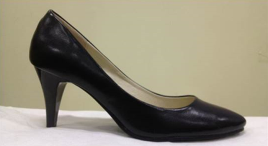
All participants wore spandex tights and also wore the same brand and model of high heels (7 cm heel and 1 cm2 bottom area) in their normal shoe size (Figure 1). Each participant performed 2 min of walking on a treadmill to be acclimated and to determine the preferred speed (walking barefoot 2.31 ± 0.31 km/h and walking in high heels 2.12 ± 0.34 km/h), after which measurements were taken while walking barefoot and then in high heels.
3. Data collection and analysis
In the experiment, data from 10 gait cycles were collected twice in an interval of 30 sec during continuous walking
1) Kinematic data
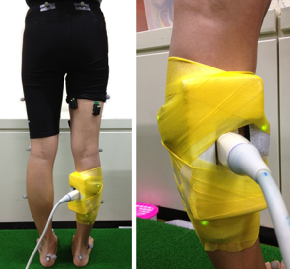
Kinematic data of the lower limb joints were collected at a frequency of 300 Hz using a 3D motion analysis system (Vicon Motion Systems, Oxford Metrics Ltd., Oxford, UK) comprised of 8 infrared cameras (Vicon MX-F20, Oxford Metric Ltd., Oxford, UK). As shown in Figure 2, round 14 mm reflective markers were attached to the left and right anterior superior iliac spine (ASIS), posterior superior iliac spine (PSIS), lateral midthigh, lateral knee epicondyle, lateral mid shank, lateral malleoli, posterior calcaneus, and second metatarsal head, which were then used to collect flexion/ extension angles of the hip, knee, and ankle joints from the sagittal plane. The joint angles were defined as flexion (+)/hyperextension (-) with 0°, where the two segments that form the hip and knee joint angles were at 180°, as the reference, while the ankle joints were defined as dor-siflexion (+)/plantar flexion (-).
2) Muscle fascicle length
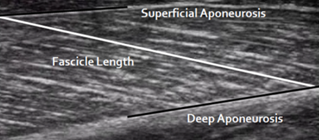
MG fascicle length was measured using a real-time ultra- sound imaging device (α10, Aloka, Japan) and a probe (Aloka 7.5 MHz UST-5712 linear-array probe, Japan). The probe was attached to the center point of the MG (Figure 2) and the ultrasound images were acquired at a frequency of 30 Hz. The acquired ultrasound images were analyzed using a free image measuring program, Image J (Wayne Rasband, National Institute of Mental Health, Bethesda, Maryland, USA), and as shown in Figure 3, muscle fascicle length was measured by connecting the superficial apo- neurosis and deep aponeurosis.
3) MTC length
The change in the length of the MG MTC was estimated by substituting the calculated joint angles into the esti-mation equation for MTC length change established by Grieve, Pheasant, & Cavanagh (1978) (Equation 1, 2, Table 1).
|
A0 |
A1 |
A2 |
|
|
Ankle |
-22.18468 |
+0.30141 |
-0.00061 |
|
Knee |
6.46251 |
-0.07987 |
+0.00011 |
4) Electromyography (EMG)
For collection of EMG data, a wireless EMG device (Trigno Wireless System, Delsys, Inc., Boston, MA, USA) was used. The surface electrodes were attached over the MG after hair was removed and the skin was cleaned with alcohol. The bandwidth of the EMG signal was set to 20~450 Hz, while the common-mode rejection ratio (CMRR) of the electrodes was set to 110 dB. The EMG signals were amp- lified by 1,000 fold and collected at a frequency of 1,500 Hz, and the signal was transmitted by a wireless trans-mitter attached to the muscle. Collected data were analyzed using EMG analysis software (EMG Works Analysis 4.0, Delsys Inc.), and after undergoing a full wave rectification process, the filtered data were used to calculate the root mean square (RMS) values using a moving window length of 50 ms and window overlap of 25 ms. From the RMS values that were calculated, peak values from each interval of the right foot's stance phase were derived for com- parison of muscle activity patterns during the gait task.
4. Interval settings
The analysis in this study focused mostly on the right foot's stance phase and the movement points according to each movement interval were as follows:
- HC : Right heel contact
- FF : Right foot flat
- MS : Mid stance
- HO : Right heel off
- TO : Right toe off
5. Statistical processing
In order to investigate the differences in muscle fascicle length according to wearing or not wearing high heels, a comparison was made using 95% confidence intervals. The differences in muscle fascicle length and muscle activity were compared using a paired t-test with a significance level of α = .05.
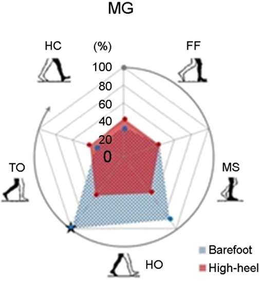
Figure 4 shows a graph of 95% confidence interval of MG fascicle length with respect to all intervals of gait cycle during walking barefoot and in high heels. In the interval from HC to HO when the foot is contacting the ground and the interval right before HC, walking in high heels, as compared to walking barefoot, showed movement with a significantly reduced range of MG fascicle length. Mean-while, the interval from HO to TO, when the right foot Table 2. The MG fascicle length change (ΔFascicle length) and muscle activation change (Δpeak EMG) during the stance phase (FF to HO) Barefoot High-heel p Mean SD Mean SD ΔFascicle length (gradient) 0.060 0.047 0.136 0.049 .001* Δpeak EMG (V) 0.030 0.011 0.016 0.019 .039* *p < .05 lifted off the ground, did not show a significant difference. In order to illustrate the amount of change in MG fascicle length, the slope of peak contraction point and peak extension point of the muscle fascicle in the gait interval from FF to HO was derived (Table 2). The results showed that the amount of change in the MG fascicle length was significantly higher, about twice as much, when walking in high heels as compared to walking barefoot (0.060 ± 0.047 vs 0.136 ± 0.049).
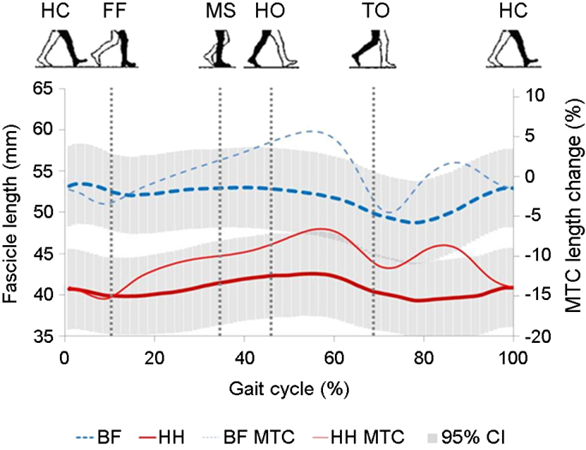
In order to illustrate the amount of change in MG fascicle length, the slope of peak contraction point and peak extension point of the muscle fascicle in the gait interval from FF to HO was derived (Table 2). The results showed that the amount of change in the MG fascicle length was significantly higher, about twice as much, when walking in high heels as compared to walking barefoot (0.060 ± 0.047 vs 0.136 ± 0.049).
|
Barefoot |
|
High-heel |
p |
|||||
|
Mean |
SD |
Mean |
SD |
|||||
|
ΔFascicle length |
0.060 |
0.047 |
|
0.136 |
0.049 |
.001* |
||
|
Δpeak EMG (V) |
0.030 |
0.011 |
|
0.016 |
0.019 |
.039* |
||
When the amount of change in peak EMG of MG between the gait cycles divided into intervals was calculated and examined after standardizing it as a ratio of peak EMG during walking barefoot (from HO to TO; Table 2, Figure 5), walking in high heels showed significantly lower amounts of change in the peak EMG of MG than walking barefoot in the interval between FF and HO of the stance phase when MTC length increases (0.030 ± 0.011 vs. 0.016 ± 0.019).
This study was conducted on healthy women who had no experience wearing high heels with the objective of investigating the pattern of change in MG length and muscle activity that appear acutely during walking on high heels. To verify this, real-time observations were made on changes in lower limb joint angles and MG fascicle length, along with muscle activity, during walking barefoot and on high heels. In this study, the results showed that in comparison to walking barefoot, walking on high heels exhibited a pattern of eccentric contraction with a signifi-cantly shorter length of MG fascicle in the interval where MTC length increased during gaiting, and the level of muscle activity was also lower. These findings are consistent with the prediction that there would be differences in MG fascicle length changes, contraction patterns, and muscle activity patterns between walking barefoot and walking on high heels.
One of the main findings in this study was the fact that throughout all intervals of the gait, the MG fascicle moved within a shorter length when wearing high heels as com-pared to the barefoot condition. Muscle fascicle length is known to have an influence on the force-length relation- ship (Lieber, Loren, & Friden, 1994; Narici, 1999). Therefore, considering the fact that when wearing high heels, as compared to being barefoot, MG is active within the range of ascending limb length that exerts less force in the force-length relationship (Kawakami, Ichinose, & Fukunaga, 1998), it can be viewed that wearing high heels causes the MG to be placed in a range of length that has relatively lower force potential from a dynamics perspective.
When walking barefoot, the ankle joints perform dorsi-flexion from when the entire foot is touching the ground (FF) to when the heel comes off (HO), and in the interval where the length of the MTC extends, the MG fascicle was maintained at a relatively consistent length. These findings are consistent with the results of prior studies (Fukunaga, Kubo, Kawakami, Fukashiro, Kanehisa, & Maganaris, 2001; Ishikawa, Komi, Grey, Lepola, & Bruggemann, 2005; Lichtwark, Bougoulias, & Wilson, 2007; and Csapo, Maganaris, Seynnes, & Narici, 2010). Meanwhile, within the same interval, muscle fascicle length increased by 2-fold when walking on high heels than when walking barefoot (Table 2). In other words, unlike concentric contractions seen when walking barefoot, a pattern of eccentric contraction with muscle activation and longer muscle length was seen when walking on high heels. With respect to the force-length relationship, the barefoot condition places the muscle in a length range that enables it to sufficiently respond to the external load exerted when pushing off the ground and moving forward, whereas wearing high heels places it in a relatively shorter length that makes it difficult to respond to external force, and as a result, it extends to a longer length that allows greater force to be expressed. Therefore, from a force-velocity relationship perspective, walking on high heels, which exhibits a pattern of eccentric contraction, may place the muscle under conditions that generate greater mechanical workload that the contractile elements must handle than walking barefoot, which exhibits a pattern of concentric contraction.
Wearing high heels resulted in decreased MG activity, which may be due to two reasons. First, the ankle joint performs plantar flexion of approximately 30° at the point of toe-off in the gait cycle. If the entire bottom of the bare foot is touching the ground, performing plantar flexion of 30° would require greater force, but when high-heels are worn, the ankle joint is already plantar flexed, and as such, it is predicted to require less muscle activity up to the toe-off point. Second, because muscle length is de- creased in comparison to being barefoot, muscle activity can be expected to be lower as well. Muscle being less active when walking with the muscle length being at a shorter length is consistent with the findings from prior studies (Lee, Matteliano, Medige, & Smiehorowski, 1987; Lee, Shieh, Matteliano, & Smiehorowski, 1990; Gregor, Smith, & Prilutsky, 2006; Lichtwark, & Wilson, 2006). Studies by Csapo, Maganaris, Seynnes, & Narici (2010) and Cronin, Barrett, & Carty (2012) on women who have worn high heels for a long time. They found that continuing to wear high heels for a long time can lead to a decrease in MG fascicle length from loss of number of muscle segments and such change may cause a chronic decrease in muscle activity. Moreover, a decrease in muscle fascicle length changes the operating range of the muscle, and as such, muscle strength expression characteristics in a force-length rela-tionship may also show chronic changes.
Wearing high heels not only causes anatomical and functional changes in muscles, but also, as shown in pre- vious studies, it could increase the stiffness of tendons by continuously exerting greater loads on the tendons. An increase in tendon stiffness may allow for efficient delivery of force through the MTC, but such morphological changes to muscle and tendons could decrease the range of motion in the ankle joints, which might present discomfort when walking in flat shoes or performing activities of daily living (Ryu, 2009; Ryu, 2010; Opila, Wagner, Schiowitz, & Chen, 1988).
This study aimed to identify the effects of wearing high¬heels on the pattern of change in MG fascicle length. The study was conducted on women without any experience of wearing high heels. The comparative analysis of walking barefoot versus in high heels showed different changing patterns in MG fascicle length and muscle activity.
In the future, for investigation of the direct effects of high heels on chronic changes in muscle fascicle length, it would be necessary for studies to include a long accli- mation period. Moreover, simultaneously measuring the changes in tendon length allows for more precise study results to be derived, while also broadening our under- standing of the interaction between muscles and tendons.
Because of wearing high heels, MG fascicle length was found to change within a relatively shorter range during the interval when the foot is touching the ground when walking, as compared to being barefoot, while the muscle fascicle exhibited a pattern of eccentric contraction in the interval where the length of the MTC increased. Moreover, the extent of change in peak EMG of MG was found to be significantly lower when walking in high heels as com- pared to walking barefoot. Based on these study results, it was determined that wearing high heels affected not only the pattern of change in muscle fascicle length and muscle activity, but that wearing high heels for a long time might result in a chronic decrease in muscle fascicle length.
References
1. Alexander, R. M. (1988). Elastic mechanisms in animal move-ment. Cambridge University Press.
Crossref
Google Scholar
2. Alexander, R. M. (2002). Tendon elasticity and muscle func- tion. Comparative Biochemistry and Physiology Part A: Molecular & Integrative Physiology, 133(4), 1001-1011.
Crossref
Google Scholar
PubMed
3. Alexander, R. M. & Bennet-Clark, H. (1977). Storage of elastic strain energy in muscle and other tissues. Nature, 265(5590), 114-117.
Crossref
Google Scholar
PubMed
4. Cavagna, G. A., Heglund, N. C. & Taylor, C. R. (1977). Mechanical work in terrestrial locomotion: two basic mechanisms for minimizing energy expenditure. American Journal of Physiology-Regulatory, Integrative and Comparative Physiology, 233(5), R243-R261.
Crossref
Google Scholar
5. Cronin, N. J., Barrett, R. S. & Carty, C. P. (2012). Long-term use of high-heeled shoes alters the neuromechanics of human walking. Journal of Applied Physiology, 112(6), 1054-1058.
Crossref
Google Scholar
PubMed
6. Csapo, R., Maganaris, C., Seynnes, O. & Narici, M. (2010). On muscle, tendon and high heels. The Journal of experimental biology, 213(15), 2582-2588.
Crossref
Google Scholar
PubMed
7. De Boer, M. D., Maganaris, C. N., Seynnes, O. R., Rennie, M. J. & Narici, M. V. (2007). Time course of muscular, neural and tendinous adaptations to 23 day uni-lateral lower-limb suspension in young men. The Journal of Physiology, 583(3), 1079-1091.
Crossref
Google Scholar
PubMed
8. Ebbeling, C. J., Hamill, J. & Crussemeyer, J. A. (1994). Lower extremity mechanics and energy cost of walking in high-heeled shoes. Journal of Orthopaedic & Sports Physical Therapy, 19(4), 190-196.
Crossref
Google Scholar
9. Fukunaga, T., Kawakami, Y., Kubo, K. & Kanehisa, H. (2002). Muscle and tendon interaction during human move- ments. Exercise and sport sciences reviews, 30(3), 106 -110.
Crossref
Google Scholar
PubMed
10. Fukunaga, T., Kubo, K., Kawakami, Y., Fukashiro, S., Kanehisa, H. & Maganaris, C. N. (2001). In vivo behaviour of human muscle tendon during walking. Proceedings of the Royal Society of London B: Biological Sciences, 268(1464), 229-233.
Crossref
Google Scholar
11. Gregor, R. J., Smith, D. W. & Prilutsky, B. I. (2006). Mech- anics of slope walking in the cat: quantification of muscle load, length change, and ankle extensor EMG patterns. Journal of Neurophysiology, 95(3), 1397-1409.
Crossref
Google Scholar
PubMed
12. Grieve, D. W., Pheasant, S. & Cavanagh, P. R (1978). Pre- diction of gastrocnemius length from knee and ankle joint posture. Biomechanics VI-A (Vol. 2, pp. 405-412): University Park Press Baltimore.
Crossref
13. Griffiths, R. (1991). Shortening of muscle fibres during stretch of the active cat medial gastrocnemius muscle: the role of tendon compliance. The Journal of Physi- ology, 436(1), 219-236.
Crossref
Google Scholar
14. Hof, A. L., Van Zandwijk, J. P. & Bobbert, A. F. (2002). Mechanics of human triceps surae muscle in walking, running and jumping. Acta Physiologica Scandinavica, 174(1), 17-30.
Crossref
Google Scholar
PubMed
15. Hong, W.-H., Lee, Y.-H., Chen, H.-C., Pei, Y.-C. & Wu, C.-Y. (2005). Influence of heel height and shoe insert on comfort perception and biomechanical performance of young female adults during walking. Foot & Ankle International, 26(12), 1042-1048.
Crossref
Google Scholar
16. Ishikawa, M., Komi, P. V., Grey, M. J., Lepola, V. & Bruggemann, G.-P. (2005). Muscle-tendon interaction and elastic energy usage in human walking. Journal of Applied Physiology, 99(2), 603-608.
Crossref
Google Scholar
PubMed
17. Ishikawa, M., Pakaslahti, J. & Komi, P. (2007). Medial gastro- cnemius muscle behavior during human running and walking. Gait & Posture, 25(3), 380-384.
Crossref
Google Scholar
PubMed
18. Kawakami, Y., Ichinose, Y. & Fukunaga, T. (1998). Archi-tectural and functional features of human triceps surae muscles during contraction. Journal of Applied Physiology, 85(2), 398-404.
Crossref
Google Scholar
PubMed
19. Lee, H. D., Han, B. R., Kim, J. S., Oh, J. H., Cho, H. Y. & Yonn, S. Y. (2015). Different behavior in muscle-tendon com- plex and fascicle length of the medial gastrocnemius muscle during gait and one-legged and two-legged vertical jumping. Korean Journal of Sports Biomechanics, 25(2), 175-182.
Crossref
20. Lee, K., Matteliano, A., Medige, J. & Smiehorowski, T. (1987). Electromyographic changes of leg muscles with heel lift: therapeutic implications. Archives of Physical Medicine and Rehabilitation, 68(5 Pt 1), 298-301.
Crossref
Google Scholar
PubMed
21. Lee, K., Shieh, J., Matteliano, A. & Smiehorowski, T. (1990). Electromyographic changes of leg muscles with heel lifts in women: therapeutic implications. Archives of Physical Medicine and Rehabilitation, 71(1), 31-33.
Crossref
Google Scholar
22. Lichtwark, G. A. & Wilson, A. (2006). Interactions between the human gastrocnemius muscle and the Achilles tendon during incline, level and decline locomotion. Journal of Experimental Biology, 209(21), 4379-4388.
Crossref
Google Scholar
23. Lichtwark, G. A., Bougoulias, K. & Wilson, A. (2007). Muscle fascicle and series elastic element length changes along the length of the human gastrocnemius during walking and running. Journal of Biomechanics, 40(1), 157-164.
Crossref
Google Scholar
24. Lieber, R. L., Loren, G. J, & Friden, J. (1994). In vivo meaure- ment of human wrist extensor muscle sarcomere length changes. Journal of Neurophysiology, 71(3), 874-881.
Crossref
Google Scholar
PubMed
25. Magnusson, S. P., Narici, M. V., Maganaris, C. N. & Kjaer, M. (2008). Human tendon behaviour and adaptation, in vivo. The Journal of Physiology, 586(1), 71-81.
Crossref
Google Scholar
PubMed
26. Narici, M. (1999). Human skeletal muscle architecture studied in vivo by non-invasive imaging techniques: functional significance and applications. Journal of Electromyography and Kinesiology, 9(2), 97-103.
Crossref
Google Scholar
27. Opila, K. A., Wagner, S. S., Schiowitz, S. & Chen, J. (1988). Postural alignment in barefoot and high-heeled stance. Spine, 13(5), 542-547.
Crossref
Google Scholar
PubMed
28. Roberts, T. J., Marsh, R. L., Weyand, P. G. & Taylor, C. R. (1997). Muscular force in running turkeys: the economy of minimizing work. Science, 275(5303), 1113-1115.
Crossref
Google Scholar
PubMed
29. Roberts, T. J. & Scales, J. A. (2002). Mechanical power output during running accelerations in wild turkeys. Journal of Experimental Biology, 205(10), 1485-1494.
Crossref
Google Scholar
PubMed
30. Roberts, T. J. & Scales, J. A. (2004). Adjusting muscle func- tion to demand: joint work during acceleration in wild turkeys. Journal of Experimental Biology, 207(23), 4165 -4174.
Crossref
Google Scholar
PubMed
31. Ryu, J. S. (2009). The temporal coordination of the lower extremity by increasing high-heel height during walking. Korean Journal of Sports Biomechanics, 19(3), 593-601.
Crossref
Google Scholar
32. Ryu, J. S. (2010). Effects of high-heeled shoe with different height on the balance during standing and walking. Korean Journal of Sports Biomechanics, 20(4), 479-486.
Crossref
Google Scholar
33. Snow, R. E. & Williams, K. R. (1994). High heeled shoes: their effect on center of mass position, posture, three¬dimensional kinematics, rearfoot motion, and ground reaction forces. Archives of Physical Medicine and Rehabilitation, 75(5), 568-576.
Crossref
Google Scholar
